

Assessment of Perfused Peripapillary Capillaries and Peripapillary Capillary Density Maps in Glaucoma Patients
- PMID: 33041444
- PMCID: PMC7520061
- DOI: 10.5455/medarh.2020.74.275-278
Assessment of Perfused Peripapillary Capillaries and Peripapillary Capillary Density Maps in Glaucoma Patients
Abstract
Introduction: Monitoring and diagnosing glaucoma until 2017 included funduscopy, IOP measurements, gonioscopy, pachymetry, as well as visual field tests, optical coherence tomography (OCT) and optical coherence tomography angiography (OCT-A). Radial peripapillary capillaries (RPC) can be observed by fluorescein angiography, as well as histologically - superficial and deep capillary layer.
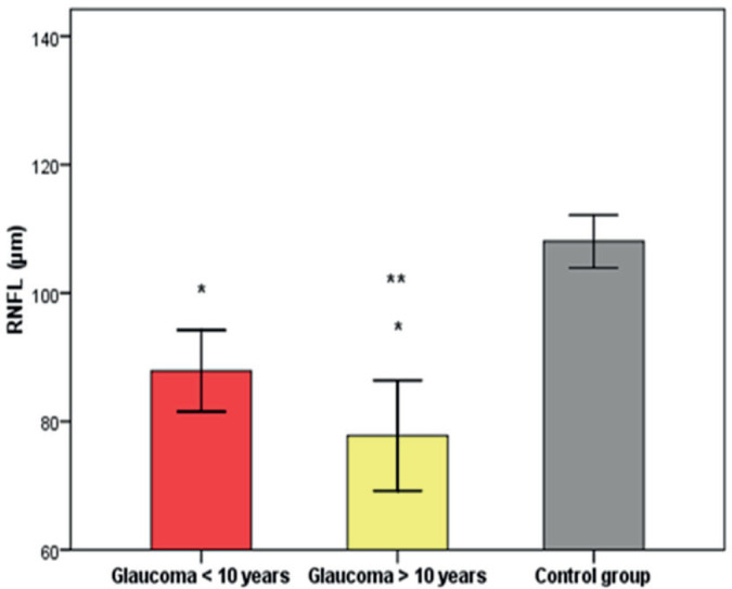
Aim: To correlate density of radial peripapillary capillary network (RPC) and retinal nerve fiber layer (RNFL) thickness in eight peripapillary segments in patients with a primary open angle glaucoma (POAG) which have the disease under 10 years of duration, over 10 years of duration and in a group of healthy patients.
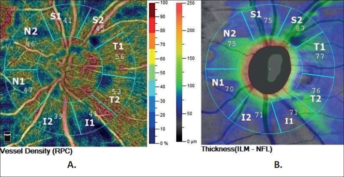
Methods: This is a cross-sectional review which included three groups of patients: POAG patients under 10 years of disease duration, over 10 years of disease duration and control group of patients. The study is performed on the commercial optical coherence tomography angiography system (AngioVue, Avanti RTVue-XR, Optovue, CA). Sectoral RPC density values, RPC maps and RNFL thickness were analyzed in three groups of patients, data was compared and correlation between parameters was examined.
Results: Mean RPC Density values in both superior segments (S1, S2) were significantly lower in patients with glaucoma over 10 years of disease duration compared to patients with glaucoma under 10 years of disease duration (p<0.05). Statistically significant positive correlation was established between RNFL thickness and RPC density in all eight peripapillary segments.
Conclusion: Analysis of radial peripapillary capillary network density on optical coherence tomography angiography may provide an earliest functional sign of progressive optic nerve disease and new insights into the pathophysiology of glaucomatous damage.
Keywords: Glaucoma; Optical Coherence Tomography Angiography; Peripapillary Capillary Density Maps; Primary Open Angle Glaucoma; Radial Peripapillary capillaries.
© 2020 Sanja Sefic, Aida Kasumovic, Ines Matoc, Tarik Halimic, Bahrija Voloder, Lejla Muhamedagic, Seldjana Catovic Delic, Irena Sesar.
Conflict of interest statement
None declared.
Figures
Similar articles
-
Quantitative Analysis of Microvasculature in Macular and Peripapillary Regions in Early Primary Open-Angle Glaucoma.Curr Eye Res. 2020 May;45(5):629-635. doi: 10.1080/02713683.2019.1676912. Epub 2019 Oct 14. Curr Eye Res. 2020. PMID: 31587582
-
Optical Coherence Tomography Angiography Analysis of Perfused Peripapillary Capillaries in Primary Open-Angle Glaucoma and Normal-Tension Glaucoma.Invest Ophthalmol Vis Sci. 2016 Jul 1;57(9):OCT611-OCT620. doi: 10.1167/iovs.15-18945. Invest Ophthalmol Vis Sci. 2016. PMID: 27742922
-
Radial Peripapillary Capillary Density Measurement Using Optical Coherence Tomography Angiography in Early Glaucoma.J Glaucoma. 2017 May;26(5):438-443. doi: 10.1097/IJG.0000000000000649. J Glaucoma. 2017. PMID: 28234680
-
The Role of Optical Coherence Tomography Angiography in Glaucoma.Semin Ophthalmol. 2024 Aug;39(6):412-423. doi: 10.1080/08820538.2024.2343049. Epub 2024 Apr 20. Semin Ophthalmol. 2024. PMID: 38643350 Review.
-
Optical Coherence Tomography Angiography in Glaucoma.J Glaucoma. 2020 Apr;29(4):312-321. doi: 10.1097/IJG.0000000000001463. J Glaucoma. 2020. PMID: 32053551 Free PMC article. Review.
Cited by
-
Predicting the impact of retinal vessel density on retinal vessel and tissue oxygenation using a theoretical model.Math Biosci. 2024 Nov;377:109292. doi: 10.1016/j.mbs.2024.109292. Epub 2024 Sep 5. Math Biosci. 2024. PMID: 39243937
-
Changes of Peripapillary Capillary Density in Patients with Vogt-Koyanagi-Harada Disease Evaluated by Optical Coherence Tomography Angiography.J Ophthalmol. 2023 Apr 17;2023:1271070. doi: 10.1155/2023/1271070. eCollection 2023. J Ophthalmol. 2023. PMID: 37102070 Free PMC article.
References
-
- Mendis KR, Balaratnasingam C, Yu P, Barry CJ, McAllister IL, Cringle SJ, Yu D. Correlation of Histologic and Clinical Images to Determine the Diagnostic Value of Fluorescein Angiography for Studying Retinal Capillary Detail. 2010;51:5864–5869. - PubMed
-
- Mase T, Ishibazawa A, Nagaoka T, Yokota H, Yoshida A. Radial Peripapillary Capillary Network Visualized Using Wide-Field Montage Optical Coherence Tomography Angiography. Invest. Ophthalmol. Vis Sci. 2016;57(12):5454. - PubMed
-
- Holló G. Vessel Density Calculated from OCT Angiography in 3 Peripapillary Sectors in Normal, Ocular Hypertensive, and Glaucoma Eyes. European Journal of Ophthalmology. 2016;26(3):42–45. - PubMed
MeSH terms
LinkOut - more resources
Full Text Sources