

Temporal Bone Chondroblastoma: a Rare Entity
- PMID: 33041452
- PMCID: PMC7520067
- DOI: 10.5455/medarh.2020.74.312-314
Temporal Bone Chondroblastoma: a Rare Entity
Abstract
Introduction: Chondroblastoma is an uncommon benign, locally destructive tumor that usually arises from epiphyses of the long bones. Temporal bone chondroblastoma is an extremely rare occurrence. Chondroblastoma arise from immature cartilage cells and it may display certain malignant features by invading surrounding structures and metastasizing to adjacent sites.
Aim: To present a case of extradural temporal bone chondroblastoma and discuss the clinical presentation, radiographic findings, histology and particularly the surgical management of the case.
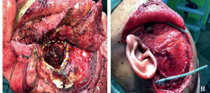
Case report: We report a case of a 31-year-old man who presented with a painless left temporal swelling and left sided hearing loss for four months. Computed tomography (CT) scan revealed an aggressive mass involving the left preauricular region with temporal mastoid bone erosion. Magnetic resonance imaging (MRI) showed an extra-axial left temporal mastoid mass pushing the left temporal lobe superiorly. The patient underwent complete excision of the temporal bone tumor. The final histopathological diagnosis was in keeping with chondroblastoma.
Conclusion: Temporal bone chondroblastoma is rare but an aggressive condition. Complete tumor resection via an appropriate approach that enables adequate exposure will lead to a favorable outcome.
Keywords: Chondroblastoma; Management; Surgical excision; Temporal bone.
© 2020 Asfa Najmi Mohamad Yusof, How Kit Thong, Tengku Mohamed Izam Tengku Kamalden.
Conflict of interest statement
The authors declare no conflicts of interest.
Figures


Similar articles
-
Chondroblastoma of the temporal bone.Acta Otolaryngol. 2011 Aug;131(8):890-5. doi: 10.3109/00016489.2011.566579. Epub 2011 Apr 19. Acta Otolaryngol. 2011. PMID: 21504272
-
Lateral skull base chondroblastoma resected with facial nerve posterior transposition.Neurochirurgie. 2017 May;63(2):88-90. doi: 10.1016/j.neuchi.2017.02.001. Epub 2017 May 11. Neurochirurgie. 2017. PMID: 28502566
-
Surface-based chondroblastoma of the tibia: a unique presentation.Skeletal Radiol. 2015 Jul;44(7):1045-50. doi: 10.1007/s00256-015-2142-4. Epub 2015 Apr 8. Skeletal Radiol. 2015. PMID: 25851832
-
Temporal bone chondroblastomas.Am J Otolaryngol. 2003 Nov-Dec;24(6):370-3. doi: 10.1016/s0196-0709(03)00084-x. Am J Otolaryngol. 2003. PMID: 14608568 Review.
-
Temporal bone chondroblastoma: a review.Neuropathology. 2005 Jun;25(2):159-64. doi: 10.1111/j.1440-1789.2005.00597.x. Neuropathology. 2005. PMID: 15875910 Review.
References
-
- Dahlin DC, Ivins JC. Benign chondroblastoma. A study of 125 cases. Cancer. 1972;30:401–413. - PubMed
-
- Codman EA. Epiphyseal chondromatous giant cell tumor of upper end of humerus. Surge Gyencol Obstet. 1931;52:543–548. - PubMed
-
- Bian LG, Sun QF, Zhao WG, Shen JK, Tirakotai W, Bertalanffy H. Temporal bone chondroblastoma: A review. Neuropathology. 2005;25:159–164. - PubMed
-
- Politi M, Consolo U, Panziera G, et al. Chondroblastoma of the temporal bone. Case report. J Craniomaxillofac Surg. 1991;19:319–322. - PubMed
Publication types
MeSH terms
LinkOut - more resources
Full Text Sources
Medical