. 2020 Jan 23;30(4):1907716.
doi: 10.1002/adfm.201907716. Epub 2019 Nov 4.

In Situ Polymerized Hollow Mesoporous Organosilica Biocatalysis Nanoreactor for Enhancing ROS-Mediated Anticancer Therapy

Affiliations

In Situ Polymerized Hollow Mesoporous Organosilica Biocatalysis Nanoreactor for Enhancing ROS-Mediated Anticancer Therapy

Ling Li et al. Adv Funct Mater. .

Abstract

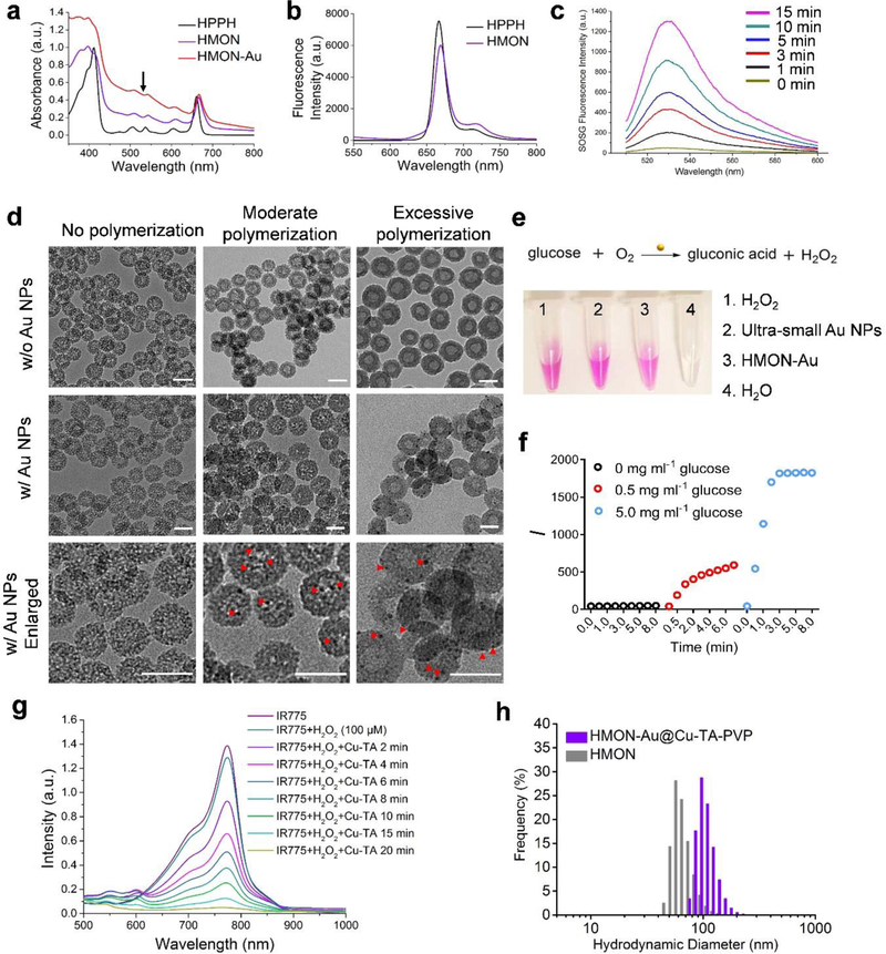
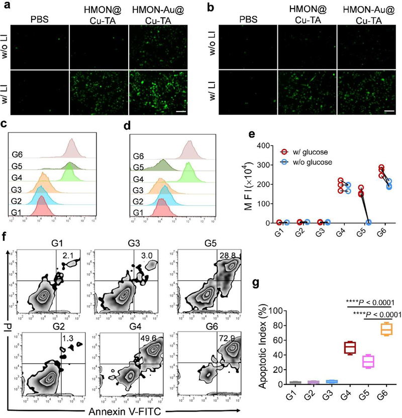
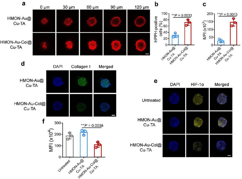
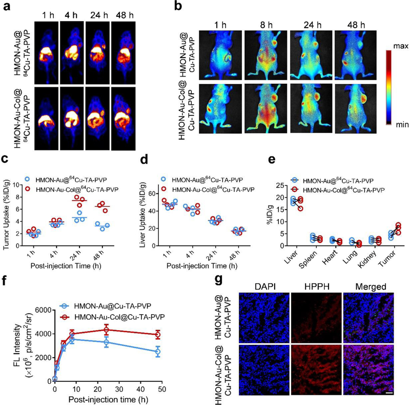
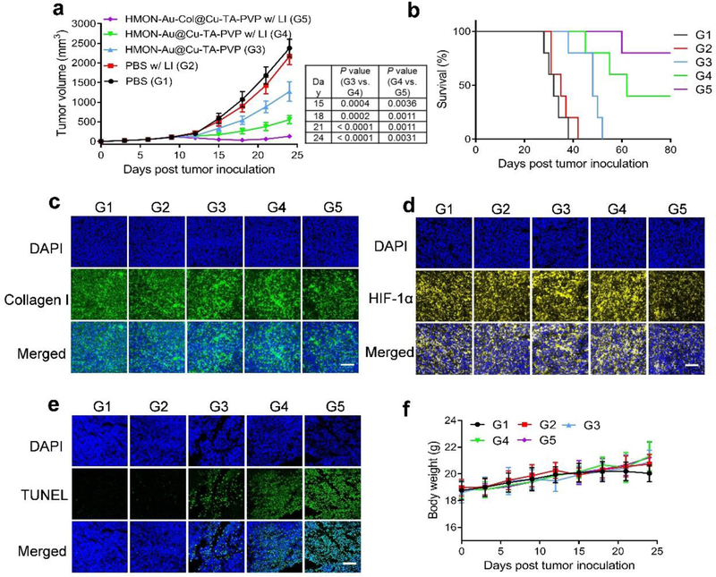
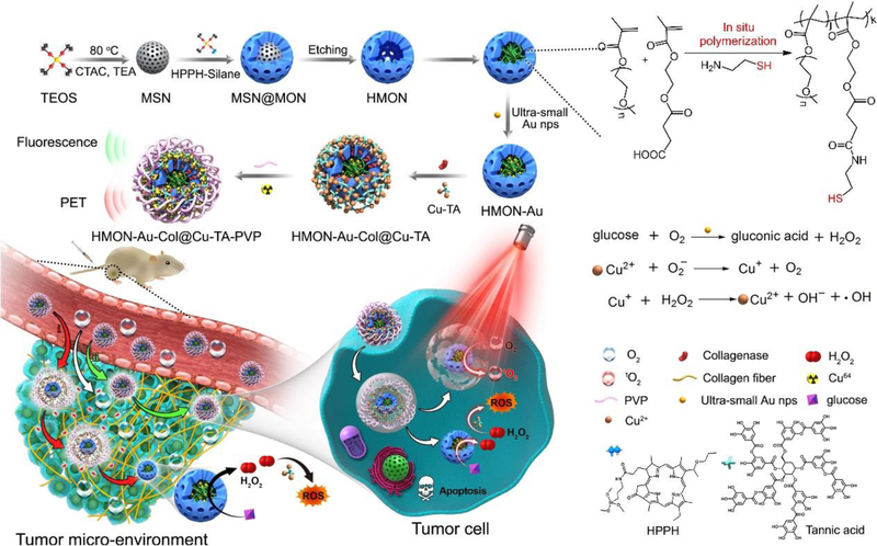
The combination of reactive oxygen species (ROS)-involved photodynamic therapy (PDT) and chemodynamic therapy (CDT) holds great promise for enhancing ROS-mediated cancer treatment. Herein, we reported an in situ polymerized hollow mesoporous organosilica nanoparticle (HMON) biocatalysis nanoreactor to integrate the synergistic effect of PDT/CDT for enhancing ROS-mediated pancreatic ductal adenocarcinoma treatment. HPPH photosensitizer was hybridized within the framework of HMON via an "in situ framework growth" approach. Then, the hollow cavity of HMONs was exploited as a nanoreactor for "in situ polymerization" to synthesize the polymer containing thiol groups, thereby enabling the immobilization of ultrasmall gold nanoparticles, which behave like glucose oxidase-like nanozyme, converting glucose into H2O2 to provide self-supplied H2O2 for CDT. Meanwhile, Cu2+-tannic acid complexes were further deposited on the surface of HMONs (HMON-Au@Cu-TA) to initiate Fenton-like reaction to covert the self-supplied H2O2 into •OH, a highly toxic ROS. Finally, collagenase (Col), which can degrade the collagen I fiber in the extracellular matrix (ECM), was loaded into HMON-Au@Cu-TA to enhance the penetration of HMONs and O2 infiltration for enhanced PDT. This study provides a good paradigm for enhancing ROS-mediated anti-tumor efficacy. Meanwhile, this research offers a new method to broaden the application of silica based nanotheranostics.

Keywords: Fenton-like reaction; chemodynamic therapy; in situ polymerization; mesoporous organosilica; ultra-small gold nanoparticle.


Figures

Figure 1.
Figure 1.
Preparation and characterization of HPPH hybridized HMONs (abbreviated as “HMON”), HMONs immobilized with the ultra-small Au NPs (HMON-Au) and HMON-Au@Cu-TA-PVP nanoparticles. (a) UV-Vis absorption of HPPH, HMON and HMON-Au. (b) Fluorescence spectra of HPPH and HMON. (c) Fluorescence spectra of the SOSG solution incubated with HMON over increased irradiation time. (d) Morphologies of HMON with no polymerization, HMON with moderate polymerization, and HMON with excessive polymerization (the upper panel, w/o Au NPs) detected by TEM; morphologies of HMON with no polymerization, HMON with moderate polymerization, and HMON with excessive polymerization incubated with the ultra-small Au NPs (the middle and lower panel, w/ Au NPs) detected by TEM. Scale bar, 50 nm. (e) The digital images of H2O2 production after indicated treatment detected by Hydrogen Peroxide Assay Kit. (f) The quantification of H2O2 production in the presence of HMON-Au at indicated concentration of glucose. (g) UV-Vis absorption of IR775, a radical indicator, after indicated treatment. (h) Size distribution of HMON and HMON-Au@Cu-TA-PVP nanoparticles measured by DLS.
Figure 2.
Figure 2.
(a-e) The detection of intracellular ROS generation after treatment using a ROS fluorescence probe DCFH-DA as the indicator. Fluorescence images (a) and flow cytometric analysis (c) of ROS generation in BxPC-3 cells after indicated treatment in the culture medium with glucose. Fluorescence images (b) and flow cytometric analysis (d) of ROS generation in BxPC-3 cells after indicated treatment in the culture medium without glucose. (e) The quantification of mean fluorescence intensity (MFI) in BxPC-3 cells after indicated treatment. (f) Flow cytometric analysis of apoptosis in BxPC-3 cells after indicated treatment. (g) The quantification of apoptotic index in BxPC-3 cells after indicated treatment. Scale bar, 100 μm. G1, PBS without LI; G2, PBS with LI; G3, HMON@Cu-TA without LI; G4, HMON@Cu-TA with LI; G5, HMON-Au@Cu-TA without LI; G6, HMON-Au@Cu-TA with LI. Data are presented as means ± s.d. Statistical significance was calculated via one-way ANOVA with Tukey post-hoc test (g).*P < 0.05, **P < 0.01, ***P < 0.001, ****P < 0.0001.
Figure 3.
Figure 3.
(a) Penetration ability of HMON-Au@Cu-TA and HMON-Au-Col@Cu-TA into 3D tumor spheroids of BxPC-3 cells. (b, c) The quantification of the percentage (b) and MFI (c) of HPPH positive cells in the 3D tumor spheroids. (d) The immunofluorescence characterization of the collagen I fiber in the 3D tumor spheroids after indicated treatment. (e) The hypoxia evaluation of 3D tumor spheroids after various treatments. Hypoxia was assessed by staining with HIF-1α (green). (f) The quantification of HIF-1α expression in 3D tumor spheroids after various treatments by flow cytometric analysis. Scale bar, 100 μm. Data are presented as means ± s.d. Statistical significance was calculated via two-tailed Student’s t-test (b, c) or one-way ANOVA with Tukey post-hoc test (f).*P < 0.05, **P < 0.01, ***P < 0.001, ****P < 0.0001.
Figure 4.
Figure 4.
In vivo distribution of HMON-Au@Cu-TA-PVP and HMON-Au-Col@Cu-TA-PVP nanoparticles in tumor-bearing mice. (a) Representative in vivo images of tumor-bearing mice at designated time point after intravenous injection with indicated formulation detected by PET imaging. (b) Representative in vivo fluorescence images of tumor-bearing mice after intravenous injection with indicated formulation. (c, d) The quantification of tumor uptake (c) and liver uptake (d) of HMON-Au@64Cu-TA-PVP or HMON-Au-Col@64Cu-TA-PVP nanoparticles over time by PET scanning at 1, 4, 24, and 48 h post injection. (n = 3/group) (e) The quantification of the PET signal intensity in the main organs of tumor-bearing mice at 48 h post injection. (f) The quantification of the fluorescence signal intensity in tumor at designated time point after indicated treatment. (g) Representative confocal laser scanning microscopy (CLSM) images of the tumor frozen section after indicated treatment. Scale bar, 100 μm.
Figure 5.
Figure 5.
The anti-tumor efficacy of synergistic PDT and CDT. (a) The average tumor growth curves after treatment. (b) Survival curves after treatment. (c-e) Immunofluorescence characterization of the collagen I fiber (c), HIF-1α (d) and TUNEL (e) in the tumor sections after treatment. (f) The body weight variation of tumor-bearing mice during treatment. Scale bar, 100 μm. G1, PBS; G2, PBS with LI; G3, HMON-Au@Cu-TA-PVP; G4, HMON-Au@Cu-TA-PVP with LI; G5, HMON-Au-Col@Cu-TA-PVP with LI. Data are presented as means ± s.d. Statistical significance was calculated via one-way ANOVA with Tukey post-hoc test (a).*P < 0.05, **P < 0.01, ***P < 0.001, ****P < 0.0001.
Scheme 1.
Scheme 1.
Schematic showing the process of preparing in situ polymerized hollow mesoporous organosilica biocatalysis nanoreactor for synergistic photodynamic (PDT)/chemodynamic (CDT) therapy.

References

    1. Chen H, Qiu Y, Ding D, Lin H, Sun W, Wang GD, Huang W, Zhang W, Lee D, Liu G, Adv. Mater 2018, 30, 1802748; - PMC - PubMed
    2. Fan W, Huang P, Chen X, Chem. Soc. Rev 2016, 45, 6488; - PubMed
    3. Wang H, Chao Y, Liu J, Zhu W, Wang G, Xu L, Liu Z, Biomaterials 2018, 181, 310; - PubMed
    4. Choi JR, Yong KW, Choi JY, Nilghaz A, Lin Y, Xu J, Lu X, Theranostics 2018, 8, 1005; - PMC - PubMed
    5. Dong Z, Feng L, Hao Y, Chen M, Gao M, Chao Y, Zhao H, Zhu W, Liu J, Liang C, Zhang Q, Liu Z, J. Am. Chem. Soc 2018, 140, 2165. - PubMed
    1. Zhou Z, Song J, Nie L, Chen X, Chem. Soc. Rev 2016, 45, 6597; - PMC - PubMed
    2. Liu C, Wang D, Zhang S, Cheng Y, Yang F, Xing Y, Xu T, Dong H, Zhang X, ACS Nano 2019, 13, 4267; - PubMed
    3. Zhu T, Shi L, Yu C, Dong Y, Qiu F, Shen L, Qian Q, Zhou G, Zhu X, Theranostics 2019, 9, 3293. - PMC - PubMed
    1. Gad H, Koolmeister T, Jemth A-S, Eshtad S, Jacques SA, Strom CE, Svensson LM, Schultz N, Lundback T, Einarsdottir BO, Nature 2017, 544, 508. - PubMed
    1. Lin L, Huang T, Song J, Ou X-Y, Wang Z, Deng H, Tian R, Liu Y, Wang J-F, Liu Y, J. Am. Chem. Soc 2019, 141, 9937; - PubMed
    2. Fan JX, Peng MY, Wang H, Zheng HR, Liu ZL, Li CX, Wang XN, Liu XH, Cheng SX, Zhang XZ, Adv. Mater 2019, 31, 1808278. - PubMed
    1. Tang Z, Liu Y, He M, Bu W, Angew. Chem. Int. Ed 2019, 58, 946; - PubMed
    2. Ranji-Burachaloo H, Gurr PA, Dunstan DE, Qiao GG, ACS Nano 2018, 12, 11819. - PubMed

LinkOut - more resources