

Long-Chain Acylcarnitines and Cardiac Excitation-Contraction Coupling: Links to Arrhythmias
- PMID: 33041874
- PMCID: PMC7518131
- DOI: 10.3389/fphys.2020.577856
Long-Chain Acylcarnitines and Cardiac Excitation-Contraction Coupling: Links to Arrhythmias
Abstract
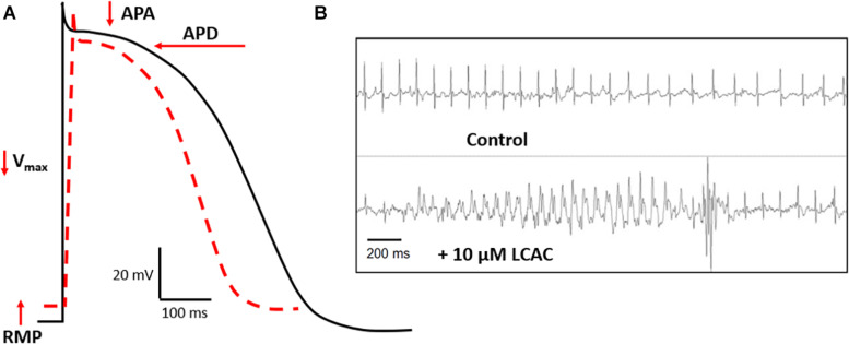
A growing number of metabolomic studies have associated high circulating levels of the amphiphilic fatty acid metabolites, long-chain acylcarnitines (LCACs), with cardiovascular disease (CVD) risk. These studies show that plasma LCAC levels can be correlated with the stage and severity of CVD and with indices of cardiac hypertrophy and ventricular function. Complementing these recent clinical associations is an extensive body of basic research that stems mostly from the twentieth century. These works, performed in cardiomyocyte and multicellular preparations from animal and cell models, highlight stereotypical derangements in cardiac electrophysiology induced by exogenous LCAC treatment that promote arrhythmic muscle behavior. In many cases, this is coupled with acute inotropic modulation; however, whether LCACs increase or decrease contractility is inconclusive. Linked to the electromechanical alterations induced by LCAC exposure is an array of effects on cardiac excitation-contraction coupling mechanisms that overload the cardiomyocyte cytosol with Na+ and Ca2+ ions. The aim of this review is to revisit this age-old literature and collate it with recent findings to provide a pathophysiological context for the growing body of metabolomic association studies that link circulating LCACs with CVD.
Keywords: arrhythmias; calcium; cardiac pathophysiology; electrophysiology; excitation-contraction coupling; long-chain acylcarnitines; metabolomics.
Copyright © 2020 Aitken-Buck, Krause, Zeller, Jones and Lamberts.
Figures


Similar articles
-
Long-chain acylcarnitine 18:1 acutely increases human atrial myocardial contractility and arrhythmia susceptibility.Am J Physiol Heart Circ Physiol. 2021 Jul 1;321(1):H162-H174. doi: 10.1152/ajpheart.00184.2021. Epub 2021 Jun 4. Am J Physiol Heart Circ Physiol. 2021. PMID: 34085842
-
Acylcarnitines--old actors auditioning for new roles in metabolic physiology.Nat Rev Endocrinol. 2015 Oct;11(10):617-25. doi: 10.1038/nrendo.2015.129. Epub 2015 Aug 25. Nat Rev Endocrinol. 2015. PMID: 26303601 Free PMC article. Review.
-
SR Ca2+-leak and disordered excitation-contraction coupling as the basis for arrhythmogenic and negative inotropic effects of acute ethanol exposure.J Mol Cell Cardiol. 2018 Mar;116:81-90. doi: 10.1016/j.yjmcc.2018.02.002. Epub 2018 Feb 3. J Mol Cell Cardiol. 2018. PMID: 29410242
-
Fibroblast growth factor-23 promotes rhythm alterations and contractile dysfunction in adult ventricular cardiomyocytes.Nephrol Dial Transplant. 2019 Nov 1;34(11):1864-1875. doi: 10.1093/ndt/gfy392. Nephrol Dial Transplant. 2019. PMID: 30629224
-
Cardiac sodium transport and excitation-contraction coupling.J Mol Cell Cardiol. 2013 Aug;61:11-9. doi: 10.1016/j.yjmcc.2013.06.003. Epub 2013 Jun 14. J Mol Cell Cardiol. 2013. PMID: 23774049 Review.
Cited by
-
Postmortem Metabolomics of Insulin Intoxications and the Potential Application to Find Hypoglycemia-Related Deaths.Metabolites. 2022 Dec 20;13(1):5. doi: 10.3390/metabo13010005. Metabolites. 2022. PMID: 36676928 Free PMC article.
-
An arrhythmogenic metabolite in atrial fibrillation.J Transl Med. 2023 Aug 24;21(1):566. doi: 10.1186/s12967-023-04420-z. J Transl Med. 2023. PMID: 37620858 Free PMC article.
-
Long-Chain Acyl-Carnitines Interfere with Mitochondrial ATP Production Leading to Cardiac Dysfunction in Zebrafish.Int J Mol Sci. 2021 Aug 6;22(16):8468. doi: 10.3390/ijms22168468. Int J Mol Sci. 2021. PMID: 34445174 Free PMC article.
-
Correlation of Serum Acylcarnitines with Clinical Presentation and Severity of Coronary Artery Disease.Biomolecules. 2022 Feb 23;12(3):354. doi: 10.3390/biom12030354. Biomolecules. 2022. PMID: 35327546 Free PMC article.
-
Metabolomic profiling in heart failure as a new tool for diagnosis and phenotyping.Sci Rep. 2025 Apr 7;15(1):11849. doi: 10.1038/s41598-025-95553-2. Sci Rep. 2025. PMID: 40195403 Free PMC article.
References
-
- Adams R. J., Cohen D. W., Gupte S., Johnson J. D., Wallick E. T., Wang T., et al. (1979a). In vitro effects of palmitylcarnitine on cardiac plasma membrane Na, K-ATPase, and sarcoplasmic reticulum Ca2+-ATPase and Ca2+ transport. J. Biol. Chem. 254 12404–12410. - PubMed
-
- Adams S. H., Hoppel C. L., Lok K. H., Zhao L., Wong S. W., Minkler P. E., et al. (2009). Plasma acylcarnitine profiles suggest incomplete long-chain fatty acid β-oxidation and altered tricarboxylic acid cycle activity in type 2 diabetic African-American women. J. Nutr. 139 1073–1081. 10.3945/jn.108.103754 - DOI - PMC - PubMed
Publication types
LinkOut - more resources
Full Text Sources
Miscellaneous