

β-arrestin 2 Is a Prognostic Factor for Survival of Ovarian Cancer Patients Upregulating Cell Proliferation
- PMID: 33042017
- PMCID: PMC7530235
- DOI: 10.3389/fendo.2020.554733
β-arrestin 2 Is a Prognostic Factor for Survival of Ovarian Cancer Patients Upregulating Cell Proliferation
Abstract
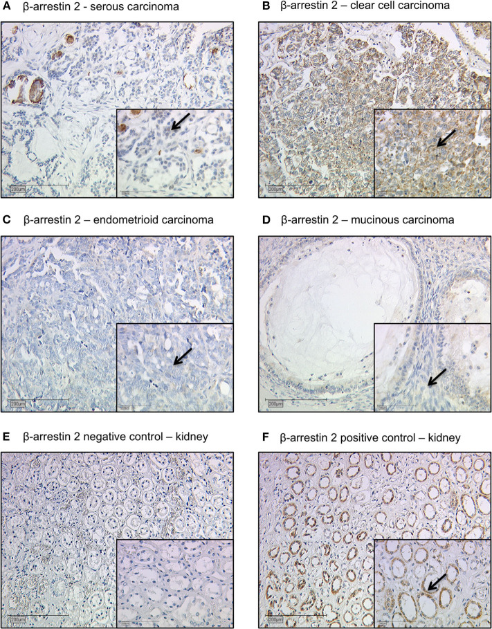
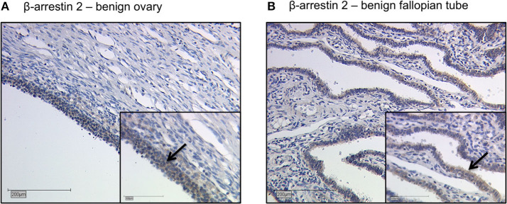
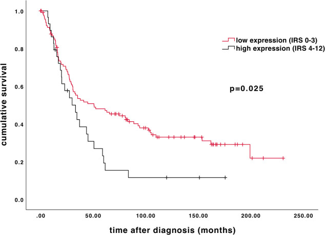
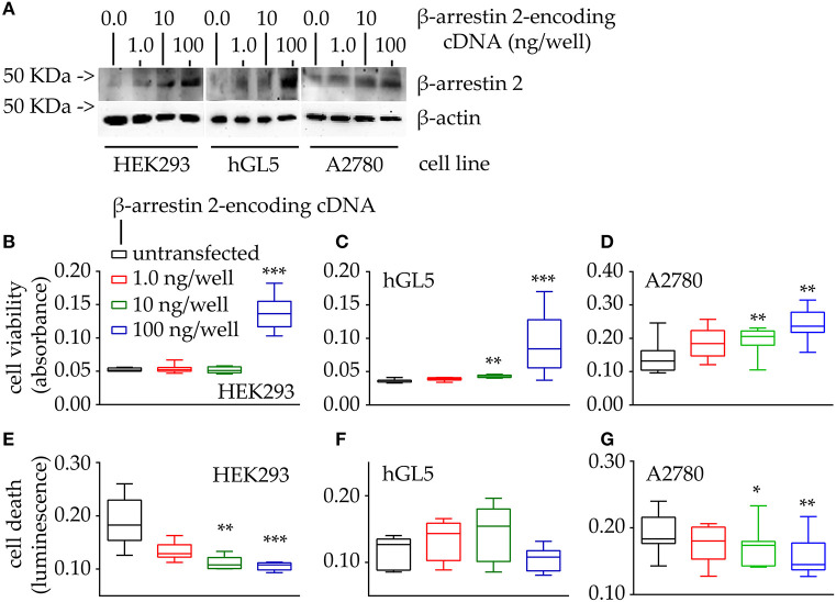
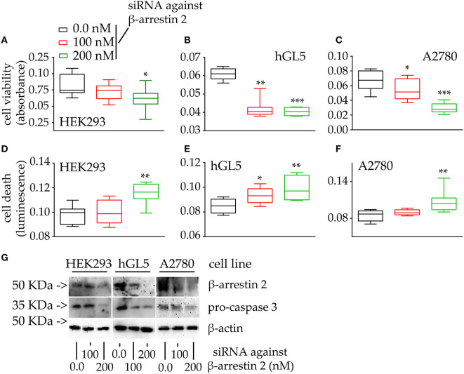
Establishing reliable prognostic factors as well as specific targets for new therapeutic approaches is an urgent requirement in advanced ovarian cancer. For several tumor entities, the ubiquitously spread scaffold protein β-arrestin 2, a multifunctional scaffold protein regulating signal transduction and internalization of activated G protein-coupled receptors (GPCRs), has been considered with rising interest for carcinogenesis. Therefore, we aimed to elucidate the prognostic impact of β-arrestin 2 and its functional role in ovarian cancer. β-arrestin 2 expression was analyzed in a subset of 156 samples of ovarian cancer patients by immunohistochemistry. Cytoplasmic expression levels were correlated with clinical as well as pathological characteristics and with prognosis. The biologic impact of β-arrestin 2 on cell proliferation and survival was evaluated, in vitro. Following transient transfection by increasing concentrations of plasmid encoding β-arrestin 2, different cell lines were evaluated in cell viability and death. β-arrestin 2 was detected in all histological ovarian cancer subtypes with highest intensity in clear cell histology. High β-arrestin 2 expression levels correlated with high-grade serous histology and the expression of the gonadotropin receptors FSHR and LHCGR, as well as the membrane estrogen receptor GPER and hCGβ. Higher cytoplasmic β-arrestin 2 expression was associated with a significantly impaired prognosis (median 29.88 vs. 50.64 months; P = 0.025). Clinical data were confirmed in transfected HEK293 cells, human immortalized granulosa cell line (hGL5) and the ovarian cancer cell line A2780 in vitro, where the induction of β-arrestin 2 cDNA expression enhanced cell viability, while the depletion of the molecule by siRNA resulted in cell death. Reflecting the role of β-arrestin 2 in modulating GPCR-induced proliferative and anti-apoptotic signals, we propose β-arrestin 2 as an important prognostic factor and also as a promising target for new therapeutic approaches in advanced ovarian cancer.
Keywords: G protein-coupled receptor; immunohistochemistry; in vitro analyses; ovarian cancer; survival analysis; β-arrestin 2.
Copyright © 2020 Czogalla, Partenheimer, Jeschke, von Schönfeldt, Mayr, Mahner, Burges, Simoni, Melli, Benevelli, Bertini, Casarini and Trillsch.
Figures
Similar articles
-
β-arrestins regulate gonadotropin receptor-mediated cell proliferation and apoptosis by controlling different FSHR or LHCGR intracellular signaling in the hGL5 cell line.Mol Cell Endocrinol. 2016 Dec 5;437:11-21. doi: 10.1016/j.mce.2016.08.005. Epub 2016 Aug 5. Mol Cell Endocrinol. 2016. PMID: 27502035
-
G Protein-Coupled Estrogen Receptor Correlates With Dkk2 Expression and Has Prognostic Impact in Ovarian Cancer Patients.Front Endocrinol (Lausanne). 2021 Feb 19;12:564002. doi: 10.3389/fendo.2021.564002. eCollection 2021. Front Endocrinol (Lausanne). 2021. PMID: 33679613 Free PMC article.
-
FOXM1 expression is significantly associated with chemotherapy resistance and adverse prognosis in non-serous epithelial ovarian cancer patients.J Exp Clin Cancer Res. 2017 May 8;36(1):63. doi: 10.1186/s13046-017-0536-y. J Exp Clin Cancer Res. 2017. PMID: 28482906 Free PMC article.
-
Correlation of the Aryl Hydrocarbon Receptor with FSHR in Ovarian Cancer Patients.Int J Mol Sci. 2019 Jun 12;20(12):2862. doi: 10.3390/ijms20122862. Int J Mol Sci. 2019. PMID: 31212758 Free PMC article.
-
Prognostic significance of USP33 in advanced colorectal cancer patients: new insights into β-arrestin-dependent ERK signaling.Oncotarget. 2016 Dec 6;7(49):81223-81240. doi: 10.18632/oncotarget.13219. Oncotarget. 2016. PMID: 27835898 Free PMC article.
Cited by
-
β-Arrestins: Structure, Function, Physiology, and Pharmacological Perspectives.Pharmacol Rev. 2023 Sep;75(5):854-884. doi: 10.1124/pharmrev.121.000302. Epub 2023 Apr 7. Pharmacol Rev. 2023. PMID: 37028945 Free PMC article. Review.
-
A diagnostic miRNA panel to detect recurrence of ovarian cancer through artificial intelligence approaches.J Cancer Res Clin Oncol. 2023 Jan;149(1):325-341. doi: 10.1007/s00432-022-04468-2. Epub 2022 Nov 15. J Cancer Res Clin Oncol. 2023. PMID: 36378340 Free PMC article.
-
A Novel Splice Variant of BCAS1 Inhibits β-Arrestin 2 to Promote the Proliferation and Migration of Glioblastoma Cells, and This Effect Was Blocked by Maackiain.Cancers (Basel). 2022 Aug 11;14(16):3890. doi: 10.3390/cancers14163890. Cancers (Basel). 2022. PMID: 36010884 Free PMC article.
-
Molecular markers predicting the progression and prognosis of human papillomavirus-induced cervical lesions to cervical cancer.J Cancer Res Clin Oncol. 2023 Aug;149(10):8077-8086. doi: 10.1007/s00432-023-04710-5. Epub 2023 Mar 31. J Cancer Res Clin Oncol. 2023. PMID: 37000261 Free PMC article. Review.
-
Using GPCRs as Molecular Beacons to Target Ovarian Cancer with Nanomedicines.Cancers (Basel). 2022 May 10;14(10):2362. doi: 10.3390/cancers14102362. Cancers (Basel). 2022. PMID: 35625966 Free PMC article. Review.
References
-
- du Bois A, Reuss A, Pujade-Lauraine E, Harter P, Ray-Coquard I, Pfisterer J. Role of surgical outcome as prognostic factor in advanced epithelial ovarian cancer: a combined exploratory analysis of 3 prospectively randomized phase 3 multicenter trials: by the arbeitsgemeinschaft gynaekologische onkologie studiengruppe ovarialkarzinom (AGO-OVAR) and the groupe d'investigateurs nationaux pour les etudes des cancers de l'Ovaire (GINECO). Cancer. (2009) 115:1234–44. 10.1002/cncr.24149 - DOI - PubMed
Publication types
MeSH terms
Substances
LinkOut - more resources
Full Text Sources
Medical