. 2020 Oct 6:17:28.
doi: 10.1186/s12979-020-00193-x. eCollection 2020.

Age-associated changes in the circulating human antibody repertoire are upregulated in autoimmunity

Affiliations

Age-associated changes in the circulating human antibody repertoire are upregulated in autoimmunity

Aaron Arvey et al. Immun Ageing. .

Abstract

Background: The immune system undergoes a myriad of changes with age. While it is known that antibody-secreting plasma and long-lived memory B cells change with age, it remains unclear how the binding profile of the circulating antibody repertoire is impacted.

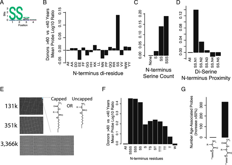
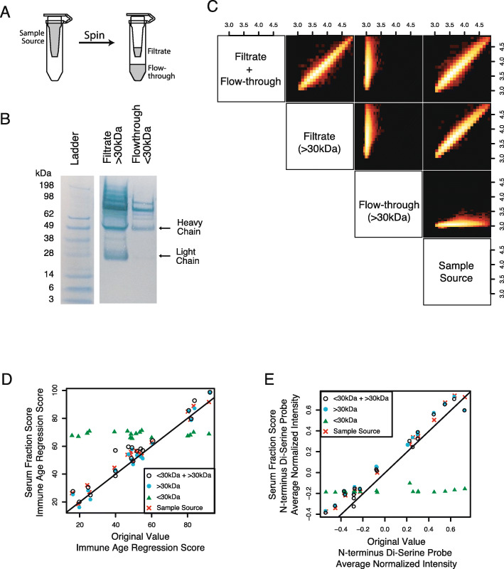
Results: To understand humoral immunity changes with respect to age, we characterized serum antibody binding to high density peptide microarrays in a diverse cohort of 1675 donors. We discovered thousands of peptides that bind antibodies in age-dependent fashion, many of which contain di-serine motifs. Peptide binding profiles were aggregated into an "immune age" by a machine learning regression model that was highly correlated with chronological age. Applying this regression model to previously-unobserved donors, we found that a donor's predicted immune age is longitudinally consistent over years, suggesting it could be a robust long-term biomarker of humoral immune ageing. Finally, we assayed serum from donors with autoimmune disease and found a significant association between "accelerated immune ageing" and autoimmune disease activity.

Conclusions: The circulating antibody repertoire has increased binding to thousands of di-serine peptide containing peptides in older donors, which can be represented as an immune age. Increased immune age is associated with autoimmune disease, acute inflammatory disease severity, and may be a broadly relevant biomarker of immune function in health, disease, and therapeutic intervention.

Keywords: Antibody binding profile; Antibody response; Auto-immune disease; Immune age; Immunosenescence; Machine learning; Peptide library.


Conflict of interest statement

Competing interestsAuthors have read the journals policy and the authors of this manuscript have the following competing interests: The authors are or were employed by HealthTell, Inc. and iCarbonX as indicated in the author’s affiliations. A patent application has been filed on the data presented. This does not alter the author’s adherence to the BMC Immunity and Ageing editorial policies.

Figures

Fig. 1
Fig. 1
Antibodies isolated from human sera show different binding profiles in older compared to younger donors. a Peptide arrays were manufactured with over 131 k diverse probes to assess IgG antibody binding. The assay workflow includes incubating donor serum sample on the peptide microarray, detecting bound IgG with a fluorophore-conjugated secondary antibody, and quantifying the fluorescent signal at each feature. A subset of four peptide features are shown along with cognate binding antibody molecules (as indicated by color). b The donor cohorts were designed to obtain diverse sampling of donor demographics, including age, BMI, sex, and geography from multisite recruitment. Combinations of age and BMI were explicitly balanced, as were other combinations of demographics. c Age (x-axis) is highly correlated with many probes’ fluorescent intensities; for example, probe XY064981 with peptide sequence SSVYDG (y-axis) fluorescent intensity and age across N = 601 donors (each datapoint) has Pearson’s correlation coefficient of r = 0.50 (p < 10− 38). d There are 100 s of peptide features that are significantly associated with older vs. younger serum donors (red points). The average peptide intensity of younger donors (x-axis) versus older donors (y-axis) shows the differential expression of all peptides. Every data point is a single peptide probe on the array. Alternative estimates of effect size and significance yield similar results (Figure S1). e Probes associated with age are highly correlated: if one age-associated peptide probe has elevated fluorescent intensity in a given donor, it is likely that fluorescence of many age-associated peptides are increased. Age-associated probes (y-axis, selected red points in Fig. 1d) are shown across all 601 donors in the cohort (x-axis). Donors are labeled by age (gray-scale legend) and probe intensities values are shown as Log10 ratio of probe in specific donor versus mean probe intensity across all donors. Hierarchical clustering was performed on donors and probes independently
Fig. 2
Fig. 2
Peptide sequence motifs in probes associated with age. a Sequence motifs in peptide probes associated with age. Peptides associated with age contain a strong N-terminus di-serine (N-di-serine) motif. Motif information content (bits, y-axis) is shown for each position (x-axis). b The N-terminus di-serine motif is much more associated with age (y-axis) than any other di-residue motif (x-axis). c The number of serine residues at the N-terminus (x-axis) is correlated with age-associated antibody binding (y-axis). d Age-associated peptide binding decreases with increased distance of di-serine from N-terminus. The starting position of di-serine residues (x-axis) relative to the N-terminus. The N-terminus is defined as N = 1. e To further characterize the peptide motifs, multiple peptide array synthesis modalities were employed (see Methods). Arrays with ~ 131 k, ~ 351 k, and ~ 3366 k probes were synthesized with peptides that had N-terminus acetyl-capping, a free N-terminus amine, or contained probes with both capped and free N-termini. f Older and younger donor sera were assayed on large microarray format with 3366 k non-control probes, which contained a broader set of peptide probes and inclusion of amino acids, including threonine and isoleucine, which were excluded in the 131 k probe microarray. The presence of multiple N-terminus serines remains the most highly significant motif, and additional serines in positions 3 and 4 may increase discrimination slightly (N = 142 probes starting with tetra-serine). Motifs including N-terminus threonine, which is biochemically similar to serine, are the second-most associated motif. Tryptophan, which is typically the ‘stickiest’ amino acid due to the aromatic indole sidechain, is shown as a negative control that is not associated with age. g Age-associated antibody binding to the di-serine N-terminus motif requires that the N-terminus be acetylated. On arrays where both acetylated and un-acetylated (uncapped free amine) probes are on present on each individual microarray, only acetylated “SS” features show age-associated binding. The number of age-associated probes with > 50% increased binding in donors > 60 yrs. vs < 40 yrs. (y-axis) is shown for uncapped free-amine probes (left) and acetyl-capped probes (right). The cutoff of 50% is representative and other cutoffs can be found in supplemental material (Figure S3G)
Fig. 3
Fig. 3
Antibody-peptide binding profiles are able to predict chronological age with high accuracy. a While the average N-di-serine probe intensity (y-axis) is highly associated with age (x-axis), the average normalized fluorescent intensity of age-associated N-di-serine probes is only moderately predictive for chronological age (Pearson’s r = 0.36). b An elastic net regression model of peptide array probe intensity data is able to predict chronological age with high accuracy on holdout examples during model training. Each data point is a single donor, showing the age of donor (x-axis) and prediction of age based on regression model of antibody binding profile (y-axis). Pearson’s correlation coefficient of r = 0.75. c The model learned from the Training Cohort is applied to the Verification Cohort. Pearson’s correlation coefficient is r = 0.74 (p < 10− 181, 95% confidence interval of [0.71, 0.76]). d The age regression residuals (y-axis) for 24 Donors (x-axis) are highly reproducible. Each donor was assayed in 16 technical replicates, which were balanced across multiple days, array manufacturing synthesis lots, secondary antibody reagent lots, and sample dilution aliquots (Methods). Each data point is a single assay for a single donor. e The age regression residual values (y-axis) are consistent across N = 16 donors that consented to regular blood draws for > 1 yr. Donors with > 5 samples over > 1 yr (N = 13) had consistent age-regression values over this time period. Data shown for all donors (lines, color indicates donor)
Fig. 4
Fig. 4
Serum antibodies are required for predicting chronological age from peptide array binding data. Furthermore, serum small molecules do not contribute to prediction of chronological age. a Schematic of column size filter. The 30 kDa filter columns can be used to separate serum molecules into flow-through fraction that contains < 30 kDa molecules and filtered fraction that contains > 30 kDa molecules. b Size filter columns are effective at depleting IgG using a 30 kDa filter, as quantified by Coomassie Blue staining. Filtrate (> 30 kDa) produces bright bands for both light and heavy chains. Flow-through (< 30 kDa) is depleted for heavy and light chain; however lower concentrations of > 30 kDa molecules can still be seen. Ladder standard and heavy/light chain weights are annotated. Image is crop edited and rotated, unedited image can be found in Figure S8. c-e Antibody purification through column filter shows that IgG is required for prediction of chronological age. Sixteen donor samples were selected to obtain coverage of chronological age regression dynamic range (Methods). These 16 samples were processed in 4 ways: (1) no processing (sample source), (2) filtered through 30 kDa column and only the filtrate (> ~ 15 kDa molecules retained; filtrate), (3) filtered through 30 kDa column and only the flow-through retained (<~ 75 kDa molecules retained; flow through), and (4) the filtrate and flow through were recombined after running through column. c Correlation between log10 peptide intensities show sample source, filtrate + flow-through, and filtrate all recapitulate original signal. In contrast, the flow-through alone, which is IgG depleted, has no correlation with original peptide-antibody binding. d In addition to raw signal being recapitulated, the machine learning regression model is recapitulated only when IgG is present. The 16 samples are plotted as machine learning regression values from the original (x-axis) and filter column-processed (y-axis). e Same as (d), but axes’ values are the di-serine peptide score rather than chronological age regression model
Fig. 5
Fig. 5
Donors with autoimmune disease have “accelerated immune ageing” as quantified by antibody binding profiles associated with higher age than subject’s chronological age at blood draw. a Longitudinal profiling of the antibody repertoire is correlated with disease activity index in donors with systemic lupus erythematosus (SLE-DAI). SLEDAI and Immune Age are shown (y-axis) relative to days since first visit (x-axis) for three donors (distinct plots). When the maximum disease activity is compared to lowest disease activity for each donor, we find that the Immune Index is higher when SLEDAI is higher (p < 0.04, paired t-test). b Age regression residuals are higher in serum from donors with autoimmune diseases. Donors with autoimmune, autoinflammatory, and phenotypically similar diseases were profiled by peptide microarray and antibody-binding prediction of age was calculated. Donors with autoimmune disease had higher antibody-based prediction of age (after correction for chronological age) than healthy control donors and donors with phenotypically similar non-autoimmune diseases. Significance was determined by a two-sided t-test comparing non-autoimmune to SLE (p < 10− 9), RA (p < 10− 5), SS (p < 10− 3). Non-autoimmune diseases included fibromyalgia (FM), osteoarthritis (OA), vascular disease (VASC), and other diseases (data not shown). Autoimmune disease profiled were Sjogren’s syndrome (SS), rheumatoid arthritis (RA), and systemic lupus erythematosus (SLE). c Donors with high autoimmune disease activity in systemic lupus erythematosus (as measured by SLEDAI), have higher age regression residuals, which suggests SLEDAI is associated with accelerated antibody binding ageing. The SLE cohort was discretized into donors that had high disease activity (> 5 SLEDAI) vs low disease activity (<=5 SLEDAI). When multiple samples were available for a given donor, the sample with highest SLEDAI was used. Donors with non-autoimmune disease had lower antibody-binding age predictions than low SLEDAI donors (p < 10− 3, two-sided t-test), who in turn had lower age predictions than high SLEDAI donors (p < 10− 5, two-sided t-test)

References

    1. Gavazzi G, Krause KH. Ageing and infection. Lancet Infect Dis. 2002;2(11):659–666. doi: 10.1016/S1473-3099(02)00437-1. - DOI - PubMed
    1. Gross PA, Hermogenes AW, Sacks HS, Lau J, Levandowski RA. The efficacy of influenza vaccine in elderly persons. A meta-analysis and review of the literature. Ann Intern Med. 1995;123(7):518–527. doi: 10.7326/0003-4819-123-7-199510010-00008. - DOI - PubMed
    1. Siegrist CA, Aspinall R. B-cell responses to vaccination at the extremes of age. Nat Rev Immunol. 2009;9(3):185–194. doi: 10.1038/nri2508. - DOI - PubMed
    1. Goronzy JJ, Weyand CM. Successful and maladaptive T cell aging. Immunity. 2017;46(3):364–378. doi: 10.1016/j.immuni.2017.03.010. - DOI - PMC - PubMed
    1. Frasca D, Blomberg BB. Effects of aging on B cell function. Curr Opin Immunol. 2009;21(4):425–430. doi: 10.1016/j.coi.2009.06.001. - DOI - PMC - PubMed

LinkOut - more resources