

Olmesartan alleviates bleomycin-mediated vascular smooth muscle cell senescence via the miR-665/SDC1 axis
- PMID: 33042414
- PMCID: PMC7540088
Olmesartan alleviates bleomycin-mediated vascular smooth muscle cell senescence via the miR-665/SDC1 axis
Abstract
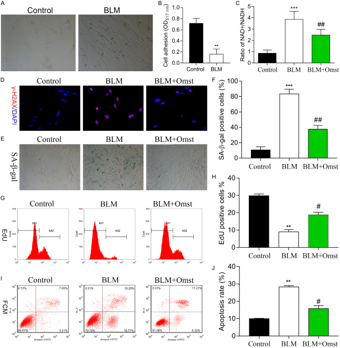
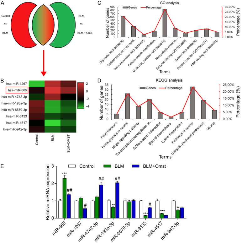
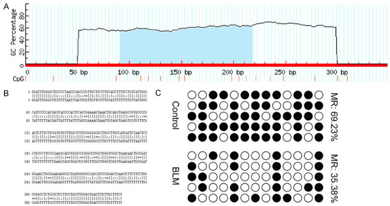
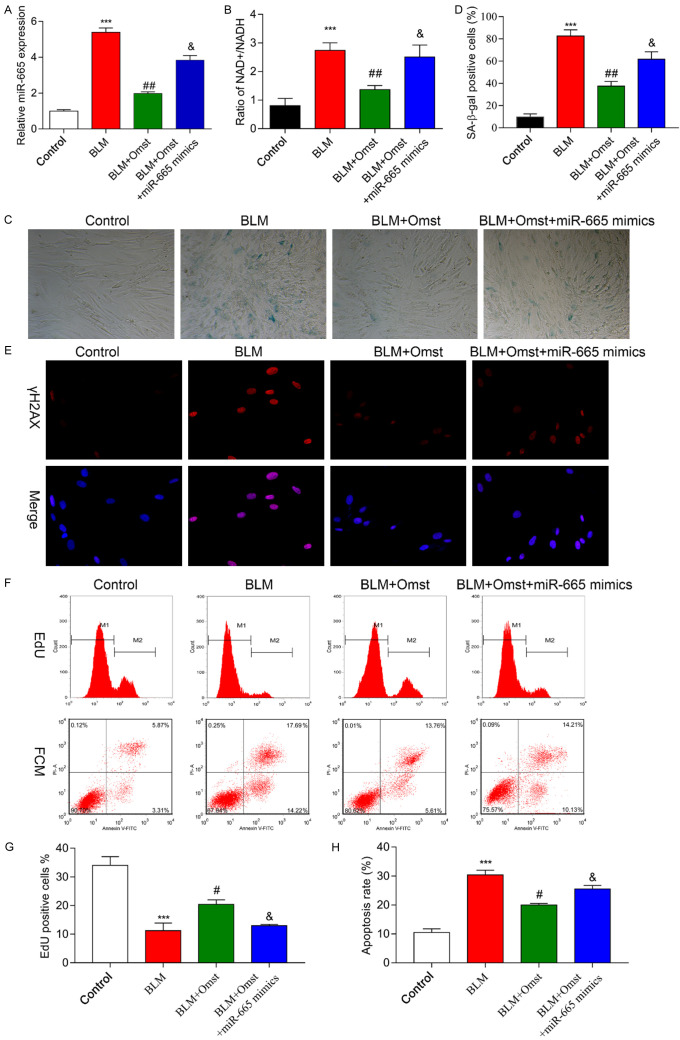
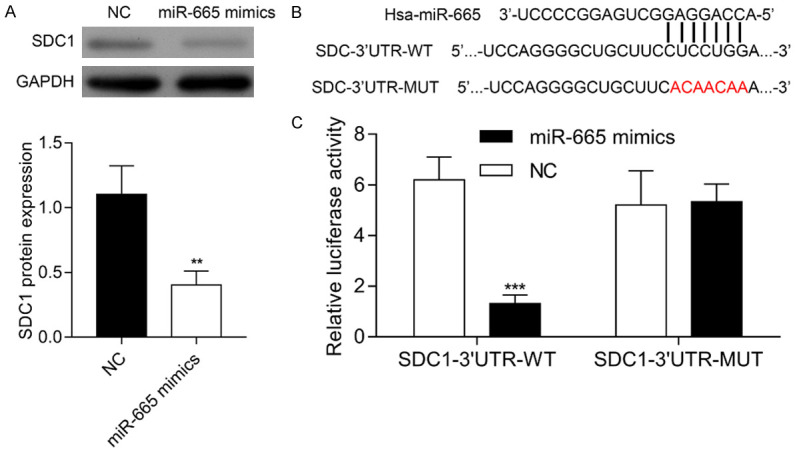
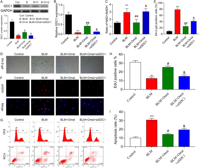
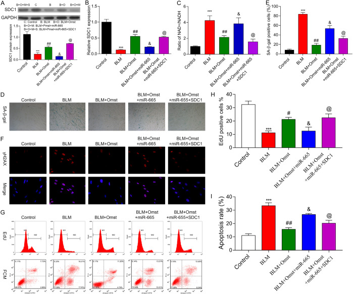
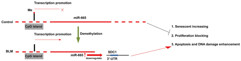
Olmesartan (OMST) is a new angiotensin II receptor antagonist recently approved by the FDA to treat cardiovascular diseases. We investigated the molecular mechanisms by which OMST regulates vascular senescence. In the present study, bleomycin (BLM) was used to induce senescence in vascular smooth muscle cells (VSMCs); after which, the cells were treated with OMST. The effects of OMST on BLM-mediated cell senescence were evaluated using cell adhesion, NAD+/NADH, and Annevin V/PI double staining assays, as well as by immunofluorescence staining of γH2AX, Edu flow cytometry, and evaluations of senescence-associated β-gal activity. Differentially expressed microRNAs (DEMs) were identified by miRNA microarray assays, and subsequently validated by quantitative real time PCR. Bisulfite sequencing PCR (BSP) was used to detect the methylation status of the miR-665 promoter. The target genes of miR-665 were predicted and confirmed using luciferase reporter assays. We found that miR-665 was upregulated in VSMCs in response to BLM-induced cellular senescence. BSP studies revealed that CpG sites in the promoter region of the miR-665 gene underwent extensive demethylation during BLM-induced cellular senescence, and there was a concomitant up-regulation of miR-665 expression. SDC1 mRNA was identified as a direct target of miR-665. Either miR-665 overexpression or SDC1 knockdown significantly reversed the effects of OMST on BLM-induced VSMC senescence. Moreover, SDC1 overexpression partially reversed the changes that occurred in cells with BLM-induced senescence caused by miR-665 overexpression. Our findings suggest that the miR-665/SDC1 axis functions as a vital modulator of VSMC senescence, and may represent a novel biological target for treating atherosclerosis.
Keywords: Atherosclerosis; MiR-665; SDC1; olmesartan; vascular smooth muscle cell senescence.
AJTR Copyright © 2020.
Conflict of interest statement
None.
Figures
Similar articles
-
Astragaloside IV alleviates senescence of vascular smooth muscle cells through activating Parkin-mediated mitophagy.Hum Cell. 2022 Nov;35(6):1684-1696. doi: 10.1007/s13577-022-00758-6. Epub 2022 Aug 4. Hum Cell. 2022. PMID: 35925474 Free PMC article.
-
MiR-665 Regulates Vascular Smooth Muscle Cell Senescence by Interacting With LncRNA GAS5/SDC1.Front Cell Dev Biol. 2021 Jul 27;9:700006. doi: 10.3389/fcell.2021.700006. eCollection 2021. Front Cell Dev Biol. 2021. PMID: 34386495 Free PMC article.
-
[Olmesartan inhibits age-associated migration and invasion of human aortic vascular smooth muscle cells by upregulating miR-3133 axis].Nan Fang Yi Ke Da Xue Xue Bao. 2020 Apr 30;40(4):499-505. doi: 10.12122/j.issn.1673-4254.2020.04.08. Nan Fang Yi Ke Da Xue Xue Bao. 2020. PMID: 32895132 Free PMC article. Chinese.
-
Myocyte Enhancer Factor 2A Regulates Hydrogen Peroxide-Induced Senescence of Vascular Smooth Muscle Cells Via microRNA-143.J Cell Physiol. 2015 Sep;230(9):2202-11. doi: 10.1002/jcp.24948. J Cell Physiol. 2015. PMID: 25655189
-
Irradiation-induced angiogenesis is associated with an MMP-9-miR-494-syndecan-1 regulatory loop in medulloblastoma cells.Oncogene. 2014 Apr 10;33(15):1922-33. doi: 10.1038/onc.2013.151. Epub 2013 Jun 3. Oncogene. 2014. PMID: 23728345
Cited by
-
Targeted inhibition of the PI3K/AKT/mTOR pathway by (+)-anthrabenzoxocinone induces cell cycle arrest, apoptosis, and autophagy in non-small cell lung cancer.Cell Mol Biol Lett. 2024 Apr 23;29(1):58. doi: 10.1186/s11658-024-00578-6. Cell Mol Biol Lett. 2024. PMID: 38649803 Free PMC article.
-
Astragaloside IV alleviates senescence of vascular smooth muscle cells through activating Parkin-mediated mitophagy.Hum Cell. 2022 Nov;35(6):1684-1696. doi: 10.1007/s13577-022-00758-6. Epub 2022 Aug 4. Hum Cell. 2022. PMID: 35925474 Free PMC article.
-
Noncoding RNAs in atherosclerosis: regulation and therapeutic potential.Mol Cell Biochem. 2024 May;479(5):1279-1295. doi: 10.1007/s11010-023-04794-0. Epub 2023 Jul 7. Mol Cell Biochem. 2024. PMID: 37418054 Free PMC article. Review.
-
ROS-mediated lysosomal membrane permeabilization and autophagy inhibition regulate bleomycin-induced cellular senescence.Autophagy. 2024 Sep;20(9):2000-2016. doi: 10.1080/15548627.2024.2353548. Epub 2024 May 18. Autophagy. 2024. PMID: 38762757 Free PMC article.
-
Correlation analysis between the expression of serum microRNA-665 and the degree of coronary artery stenosis and major adverse cardiovascular events in patients with acute myocardial infarction.J Cardiothorac Surg. 2024 Sep 23;19(1):543. doi: 10.1186/s13019-024-02998-z. J Cardiothorac Surg. 2024. PMID: 39307907 Free PMC article.
References
-
- Wang J, Uryga AK, Reinhold J, Figg N, Baker L, Finigan A, Gray K, Kumar S, Clarke M, Bennett M. Vascular smooth muscle cell senescence promotes atherosclerosis and features of plaque vulnerability. Circulation. 2015;132:1909–1919. - PubMed
-
- Liu T, Xu J, Guo JL, Lin CY, Luo WM, Yuan Y, Liu H, Zhang J. YAP1 up-regulation inhibits apoptosis of aortic dissection vascular smooth muscle cells. Eur Rev Med Pharmacol Sci. 2017;21:4632–4639. - PubMed
-
- Luo Z, Xu W, Ma S, Qiao H, Gao L, Zhang R, Yang B, Qiu Y, Chen J, Zhang M, Tao B, Cao F, Wang Y. Moderate autophagy inhibits vascular smooth muscle cell senescence to stabilize progressed atherosclerotic plaque via the mTORC1/ULK1/ATG13 signal pathway. Oxid Med Cell Longev. 2017;2017:3018190. - PMC - PubMed
LinkOut - more resources
Full Text Sources
Other Literature Sources
Research Materials
Miscellaneous