

Renal denervation mitigates atherosclerosis in ApoE-/- mice via the suppression of inflammation
- PMID: 33042425
- PMCID: PMC7540133
Renal denervation mitigates atherosclerosis in ApoE-/- mice via the suppression of inflammation
Abstract
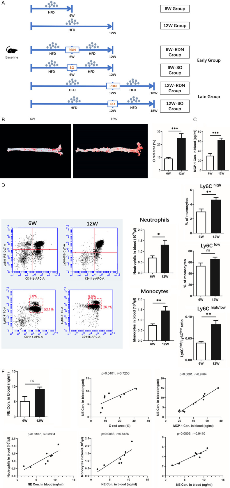
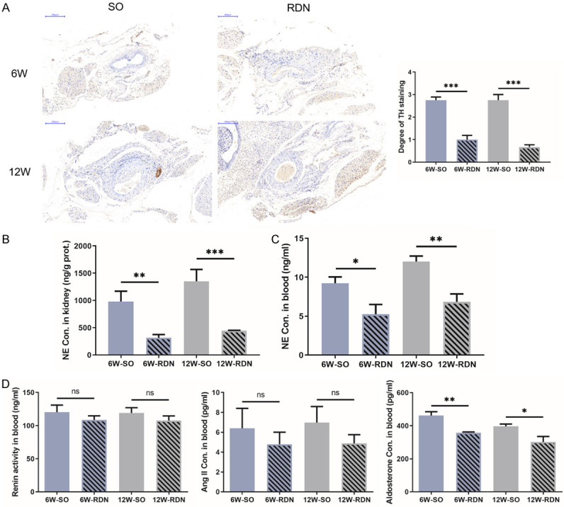
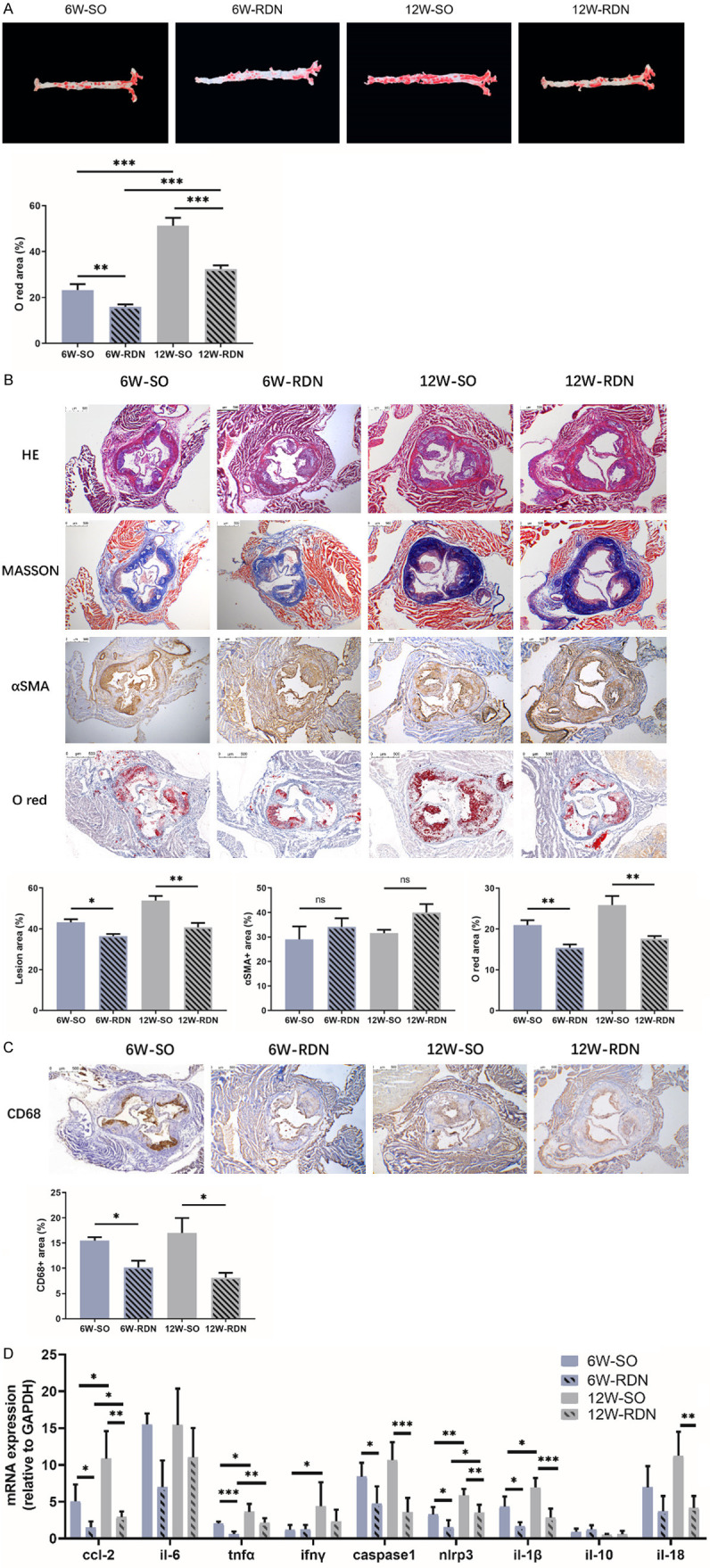
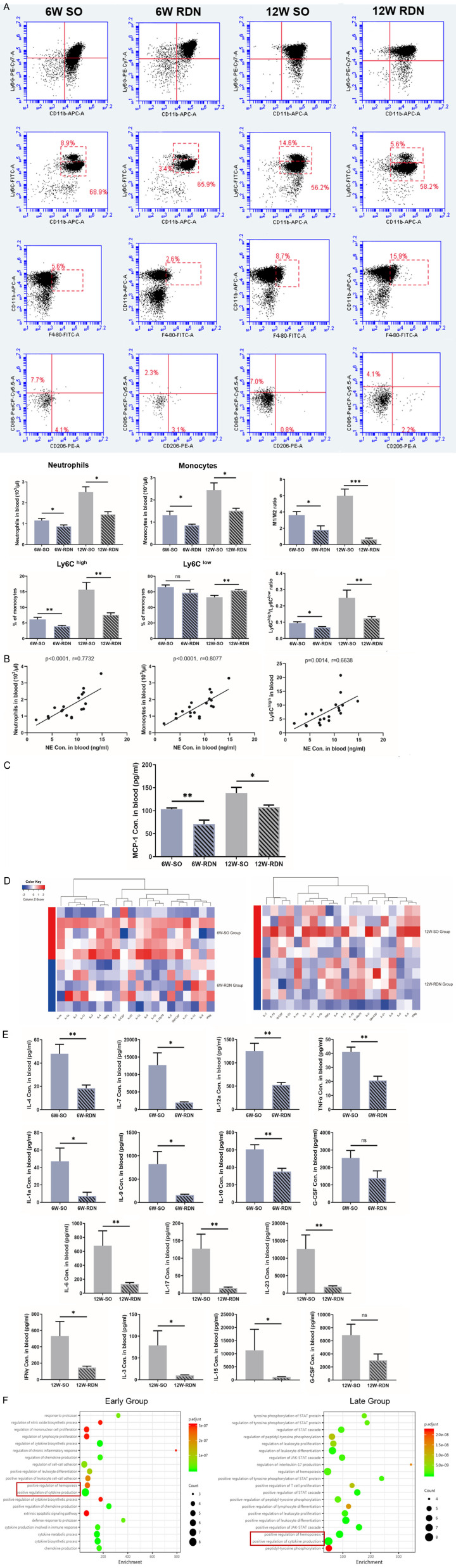
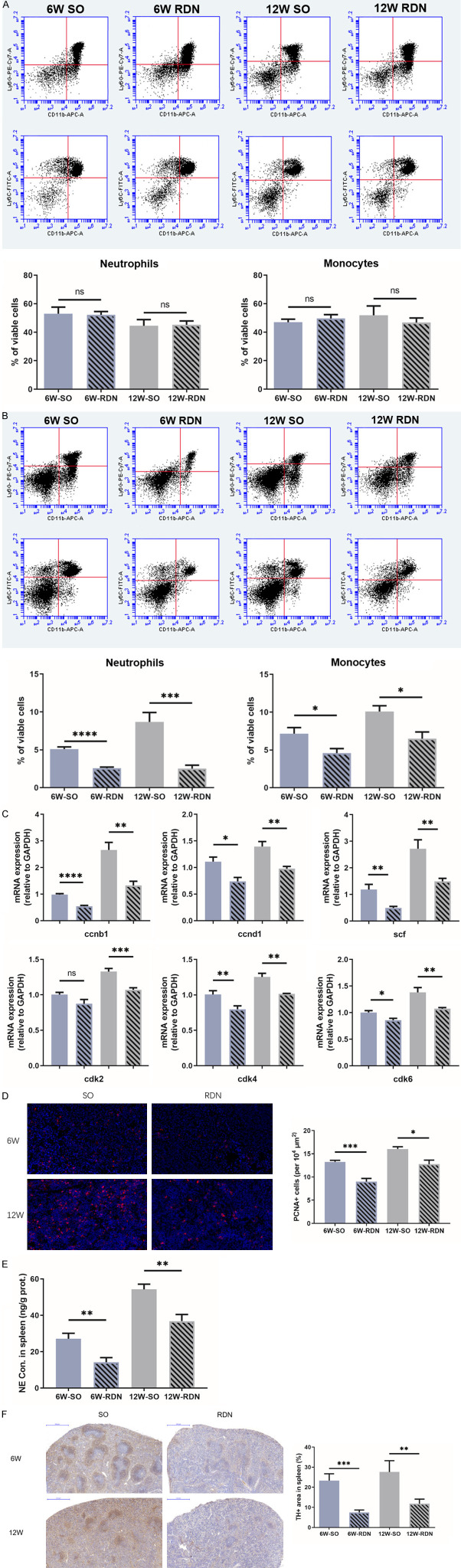
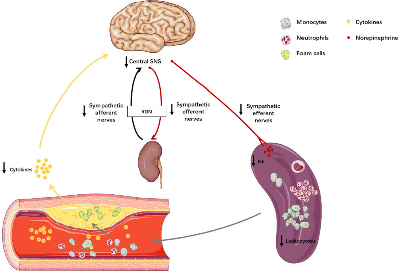
Atherosclerosis is a chronic pathological process characterized by the accumulation of inflammation. Overactivation of the sympathetic nervous system accelerates the progression of atherosclerosis. Renal denervation (RDN) reduces the activity of the sympathetic nerve system (SNS) by disrupting sympathetic nerves surrounding renal arteries. We sought to determine whether RDN could mitigate atherosclerosis through the suppression of inflammation. First, we investigated the correlation between plasma norepinephrine concentrations and circulatory inflammation in the progression of atherosclerosis. Then, forty ApoE-/- mice underwent renal denervation or a sham operation after 6 weeks or 12 weeks of feeding with a high-fat diet. The effects of RDN on atherosclerosis in mice were explored. In the development of atherosclerosis, positive correlations were found between SNS activation and the accumulation of circulatory myeloid cells and inflammatory cytokines. In the second part of the study, inhibition of the increase in plaque size was found in both RDN groups compared with that in the sham operation (SO) groups (P<0.05), and RDN also ameliorated inflammation in plaques. Furthermore, RDN attenuated the accumulation of circulating neutrophils and monocytes (P<0.05), which is associated with a significant reduction in levels of several circulating inflammatory cytokines related to hemopoiesis (P<0.05). Flow cytometry analysis revealed comparable levels of neutrophils and monocytes in the bone marrow between all four groups. However, RDN decreased the production and proportions of neutrophils and monocytes in the spleen and reduced splenic sympathetic activity (P<0.05). In summary, our study reveals a novel link between SNS activity and inflammation in atherosclerosis and identifies RDN as a potential anti-inflammatory therapeutic strategy for the treatment of atherosclerosis by restricting the production of splenic immune cells.
Keywords: Renal denervation; atherosclerosis; inflammation; sympathetic nerve system.
AJTR Copyright © 2020.
Conflict of interest statement
None.
Figures
Similar articles
-
Renal denervation restrains the inflammatory response in myocardial ischemia-reperfusion injury.Basic Res Cardiol. 2020 Jan 13;115(2):15. doi: 10.1007/s00395-020-0776-4. Basic Res Cardiol. 2020. PMID: 31932910
-
Targeting mitochondria-inflammation circle by renal denervation reduces atheroprone endothelial phenotypes and atherosclerosis.Redox Biol. 2021 Nov;47:102156. doi: 10.1016/j.redox.2021.102156. Epub 2021 Sep 29. Redox Biol. 2021. PMID: 34607159 Free PMC article.
-
Modulation of the sympathetic nervous system by renal denervation prevents reduction of aortic distensibility in atherosclerosis prone ApoE-deficient rats.J Transl Med. 2016 Jun 8;14(1):167. doi: 10.1186/s12967-016-0914-9. J Transl Med. 2016. PMID: 27277003 Free PMC article.
-
Renal Nerve Stimulation as Procedural End Point for Renal Sympathetic Denervation.Curr Hypertens Rep. 2018 Mar 19;20(3):24. doi: 10.1007/s11906-018-0821-y. Curr Hypertens Rep. 2018. PMID: 29556850 Review.
-
Selective vs. Global Renal Denervation: a Case for Less Is More.Curr Hypertens Rep. 2018 May 1;20(5):37. doi: 10.1007/s11906-018-0838-2. Curr Hypertens Rep. 2018. PMID: 29717380 Review.
Cited by
-
Favorable effect of renal denervation on elevated renal vascular resistance in patients with resistant hypertension and type 2 diabetes mellitus.Front Cardiovasc Med. 2022 Dec 14;9:1010546. doi: 10.3389/fcvm.2022.1010546. eCollection 2022. Front Cardiovasc Med. 2022. PMID: 36601066 Free PMC article.
-
Empagliflozin protects against atherosclerosis progression by modulating lipid profiles and sympathetic activity.Lipids Health Dis. 2021 Jan 12;20(1):5. doi: 10.1186/s12944-021-01430-y. Lipids Health Dis. 2021. PMID: 33436015 Free PMC article.
-
Enhanced Peripheral Chemoreflex Drive Is Associated with Cardiorespiratory Disorders in Mice with Coronary Heart Disease.Adv Exp Med Biol. 2023;1427:99-106. doi: 10.1007/978-3-031-32371-3_11. Adv Exp Med Biol. 2023. PMID: 37322340
-
Sympathetic Nervous System and Atherosclerosis.Int J Mol Sci. 2023 Aug 23;24(17):13132. doi: 10.3390/ijms241713132. Int J Mol Sci. 2023. PMID: 37685939 Free PMC article. Review.
-
The Effect of Renal Denervation on Cardiac Diastolic Function in Patients with Hypertension and Paroxysmal Atrial Fibrillation.Evid Based Complement Alternat Med. 2022 May 28;2022:2268591. doi: 10.1155/2022/2268591. eCollection 2022. Evid Based Complement Alternat Med. 2022. Retraction in: Evid Based Complement Alternat Med. 2023 Jun 21;2023:9790680. doi: 10.1155/2023/9790680. PMID: 35668773 Free PMC article. Retracted.
References
-
- Nathan C, Ding A. Nonresolving inflammation. Cell. 2010;140:871–882. - PubMed
-
- Dutta P, Courties G, Wei Y, Leuschner F, Gorbatov R, Robbins CS, Iwamoto Y, Thompson B, Carlson AL, Heidt T, Majmudar MD, Lasitschka F, Etzrodt M, Waterman P, Waring MT, Chicoine AT, van der Laan AM, Niessen HW, Piek JJ, Rubin BB, Butany J, Stone JR, Katus HA, Murphy SA, Morrow DA, Sabatine MS, Vinegoni C, Moskowitz MA, Pittet MJ, Libby P, Lin CP, Swirski FK, Weissleder R, Nahrendorf M. Myocardial infarction accelerates atherosclerosis. Nature. 2012;487:325–329. - PMC - PubMed
LinkOut - more resources
Full Text Sources
Miscellaneous