Central Nervous System Vasculitis Secondary to Sarcoidosis: A Rare Case of Lupus Pernio With Complete Occlusion of Right Internal Carotid Artery
- PMID: 33042710
- PMCID: PMC7538030
- DOI: 10.7759/cureus.10274
Central Nervous System Vasculitis Secondary to Sarcoidosis: A Rare Case of Lupus Pernio With Complete Occlusion of Right Internal Carotid Artery
Abstract
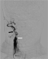
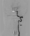
Sarcoidosis is a systemic inflammatory disorder resulting from an inappropriate immune response to ubiquitous environmental stimuli. It has a predilection for African Americans and people of Northern European countries. The classic histology is that of a non-caseating granuloma. Central nervous system involvement is a rare occurrence in sarcoidosis and even in this manifestation, the presence of vasculitis is comparatively uncommon. We present a case of a 35-year-old female, who presented with complaints of persistent headache of moderate intensity and had a violaceous plaque on nose, being treated by a dermatologist. The patient on further workup had mildly raised proteins on cerebrospinal fluid analysis. MRI brain showed multiple foci in bilateral frontoparietal regions and centrum semiovale, while digital subtraction angiography brain depicted vasculitis of small vessels of brain and complete occlusion of right internal carotid artery at its origin. Biopsy of lesion on nose was performed that showed chronic granulomatous inflammation. A diagnosis of brain vasculitis secondary to sarcoidosis was made. The patient was treated with plasmapheresis and pulse steroid therapy initially, and later on with cyclophosphamide and azathioprine. This resulted in resolution of headache and nose lesion.
Keywords: central nervous system vasculitis; digital subtraction angiography; internal carotid artery occlusion; lupus pernio; neurosarcoidosis; non-caseating granuloma; sarcoidosis.
Copyright © 2020, Arif et al.
Conflict of interest statement
The authors have declared that no competing interests exist.
Figures


Similar articles
-
Lupus pernio without systemic involvement.Indian Dermatol Online J. 2013 Oct;4(4):314-7. doi: 10.4103/2229-5178.120656. Indian Dermatol Online J. 2013. PMID: 24350015 Free PMC article.
-
A case of chronic sarcoidosis presenting with lupus pernio.Postgrad Med. 2020 Aug;132(6):532-535. doi: 10.1080/00325481.2020.1733863. Epub 2020 Feb 27. Postgrad Med. 2020. PMID: 32105165
-
Lupus Pernio.2023 Jul 10. In: StatPearls [Internet]. Treasure Island (FL): StatPearls Publishing; 2025 Jan–. 2023 Jul 10. In: StatPearls [Internet]. Treasure Island (FL): StatPearls Publishing; 2025 Jan–. PMID: 30725653 Free Books & Documents.
-
Lupus pernio.Lupus. 1992 May;1(3):129-31. doi: 10.1177/096120339200100302. Lupus. 1992. PMID: 1301972 Review.
-
SEVERE PANUVEITIS, RETINAL VASCULITIS, AND OPTIC DISK GRANULOMA SECONDARY TO SARCOIDOSIS.Retin Cases Brief Rep. 2016 Fall;10(4):341-4. doi: 10.1097/ICB.0000000000000254. Retin Cases Brief Rep. 2016. PMID: 26650564 Free PMC article. Review.
Cited by
-
Cerebral vasculitis related to neurosarcoidosis: a case series and systematic literature review.J Neurol. 2025 Jan 15;272(2):135. doi: 10.1007/s00415-024-12868-2. J Neurol. 2025. PMID: 39812656 Free PMC article.
-
Carotid dissection and central serous chorioretinopathy related to sarcoidosis-antiphospholipid syndrome: a case report.Rom J Ophthalmol. 2022 Apr-Jun;66(2):193-197. doi: 10.22336/rjo.2022.38. Rom J Ophthalmol. 2022. PMID: 35935073 Free PMC article.
References
-
- Sarcoidosis. Iannuzzi MC, Rybicki BA, Teirstein AS. N Engl J Med. 2007;357:2153–2165. - PubMed
-
- Imaging of neurosarcoidosis: common, uncommon, and rare. Bathla G, Singh AK, Policeni B, Agarwal A, Case B. Clin Radiol. 2016;71:96–106. - PubMed
-
- Four decades of necrotizing sarcoid granulomatosis: what do we know now? Rosen Y. Arch Pathol Lab Med. 2015;139:252–262. - PubMed
-
- Neurosarcoidosis. Burns TM. Arch Neurol. 2003;60:1166–1168. - PubMed
-
- Neurosarcoidosis: a study of 30 new cases. Joseph FG, Scolding NJ. J Neurol Neurosurg Psychiatry. 2009;80:297–304. - PubMed
Publication types
LinkOut - more resources
Full Text Sources
