. 2020 Aug 4;7(19):2000383.
doi: 10.1002/advs.202000383. eCollection 2020 Oct.

Adverse Impact of Heavy Metals on Bone Cells and Bone Metabolism Dependently and Independently through Anemia

Affiliations

Adverse Impact of Heavy Metals on Bone Cells and Bone Metabolism Dependently and Independently through Anemia

Shuping Zhang et al. Adv Sci (Weinh). .

Abstract

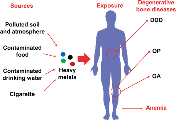
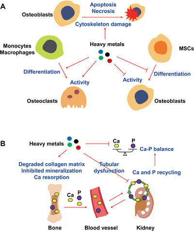
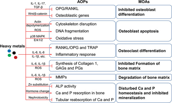
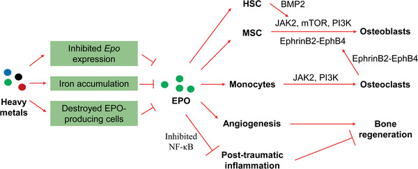
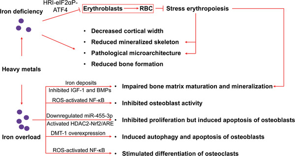
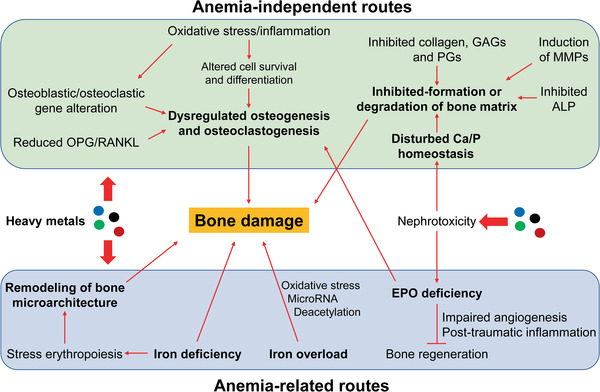
Mounting evidence is revealing that heavy metals can incur disordered bone homeostasis, leading to the development of degenerative bone diseases, including osteoporosis, osteoarthritis, degenerative disk disease, and osteomalacia. Meanwhile, heavy metal-induced anemia has been found to be intertwined with degenerative bone diseases. However, the relationship and interplay among these adverse outcomes remain elusive. Thus, it is of importance to shed light on the modes of action (MOAs) and adverse outcome pathways (AOPs) responsible for degenerative bone diseases and anemia under exposure to heavy metals. In the current Review, the epidemiological and experimental findings are recapitulated to interrogate the contributions of heavy metals to degenerative bone disease development which may be attributable dependently and independently to anemia. A few likely mechanisms are postulated for anemia-independent degenerative bone diseases, including dysregulated osteogenesis and osteoblastogenesis, imbalanced bone formation and resorption, and disturbed homeostasis of essential trace elements. By contrast, remodeled bone microarchitecture, inhibited erythropoietin production, and disordered iron homeostasis are speculated to account for anemia-associated degenerative bone disorders upon heavy metal exposure. Together, this Review aims to elaborate available literature to fill in the knowledge gaps in understanding the detrimental effects of heavy metals on bone cells and bone homeostasis through different perspectives.

Keywords: adverse outcome pathways; anemia; degenerative bone diseases; heavy metals; mode of actions.


Conflict of interest statement

The authors declare no conflict of interest.

Figures

Figure 1
Figure 1
Degenerative bone diseases and anemia under exposure to heavy metals. Polluted soil and atmosphere, contaminated food, drinking water, and cigarette smoke are the main sources for heavy metal exposure. Mounting evidence reveals that heavy metal exposure increases the risk of anemia and the development of degenerative bone diseases, e.g., osteoporosis (OP), osteoarthritis (OA) and degenerative disk disease (DDD).
Figure 2
Figure 2
Imbalanced bone formation and resorption, and disturbed homeostasis of essential trace elements in the bone under exposure to heavy metals. A) Under exposure to heavy metals, bone formation is suppressed by inhibiting the activity of osteoblasts and the differentiation of mesenchymal stem cells (MSCs) into osteoblasts. Cytoskeleton damage and cell death of osteoblasts through the induction of apoptosis or necrosis are also involved in the suppression of bone formation. Additionally, bone resorption is enhanced by increasing the activity of osteoblasts and promoting the differentiation of monocytes/macrophages into osteoclasts. B) Degradation of the collagen matrix, inhibition of mineralization and increase of calcium (Ca) resorption are induced by heavy metals, leading to the excretion of Ca and phosphorus (P). Heavy metal‐induced tubular dysfunction inhibits the resorption and recycling of Ca and P in the kidney. These outcomes ultimately induce the loss of Ca and P.
Figure 3
Figure 3
MOAs and AOPs underlying anemia‐independent detrimental effects on bone upon heavy metal exposure. Dysregulated osteogenesis and osteoclastogenesis are largely induced by alterations in the OPG/RANKL ratio and the altered expression of osteoblastic and osteoclastic genes. Oxidative stress and/or inflammation not only alter the expression of osteoblastic and osteoclastic genes, but also induce direct effects on bone. Additionally, oxidative stress and/or inflammation are actively involved in inhibiting the formation and enhancing the degradation of the extracellular bone matrix. Suppressed ALP activity and disturbed Ca and P homeostasis contribute to inhibited bone mineralization. MOAs, modes of action; AOPs, adverse outcome pathways; OPG, osteoprotegerin; RANKL, receptor activator of NF‐κB ligand; TRAP, tartrate‐resistant acid phosphatase; MMPs, matrix metalloproteinases; GAGs, glycosaminoglycans; PGs, proteoglycans; ALP, alkaline phosphatase.
Figure 4
Figure 4
Roles of hypo‐produced EPO in the anemia‐related detrimental effects on the bone under heavy metal exposure. The renal hypoproduction of EPO is induced by heavy metals. EPO deficiency impairs osteogenesis by downregulating several key signaling pathways in osteoblasts and/or osteoblasts/HSCs. Upon EPO deficiency, post‐traumatic inflammation is enhanced, and angiogenesis is impaired during bone healing. Additionally, EPO deficiency fails to induce vascular endothelial growth factor (VEGF) expression and increase peripheral endothelial progenitor cells. EPO, erythropoietin; HSC, hematopoietic stem cell; MSC, mesenchymal stem cell; JAK2, Janus kinase 2; mTOR, mechanistic target of rapamycin; PI3K, phosphoinositide 3‐kinase; BMP2, bone morphogenetic protein 2.
Figure 5
Figure 5
Mechanisms underlying the bone damage mediated by iron deficiency and iron overload. Both iron deficiency and iron overload can cause detrimental effects on bone metabolism upon heavy metal exposure. The HRI‐eIF2αP‐ATF4 signaling pathway might be involved in bone damage under iron deficiency via regulation of stress erythropoiesis. Dysregulated hepcidin, BMP, and IGF signaling pathways could be involved in bone damage observed under iron overload. The induction of oxidative stress, dysregulated microRNA and histone deacetylation by iron overload could contribute to dysregulated osteogenesis and osteoblastogenesis. RBC, red blood cell; ROS, reactive oxygen species; DMT‐1, divalent metal transporter 1; IGF‐1, insulin‐like growth factor 1; BMPs, bone morphogenetic proteins.
Figure 6
Figure 6
A global view of MOAs and AOPs underlying heavy metal‐induced bone damage. Various modes of action (MOAs) and adverse outcome pathways (AOPs) are involved in heavy metal‐induced bone damage through anemia‐independent and ‐related routes. The MOAs involved in the anemia‐independent routes mainly include dysregulated osteogenesis and osteoblastogenesis, inhibited formation and enhanced degradation of the extracellular bone matrix, disturbed Ca/P homeostasis, and inhibited mineralization. Altered OPG/RANKL ratio and altered expression of osteoblastic and osteoclastic genes are key AOPs underlying dysregulated osteogenesis and osteoblastogenesis. Nephrotoxicity is the main mechanism for the disordered Ca/P homeostasis, contributing to damage in the bone matrix together with altered levels of other matrix essentials and ALP. Regarding anemia‐related routes, stress erythropoiesis‐driven remodeling of bone microarchitecture, renal EPO hypoproduction, and disordered iron homeostasis are the main MOAs. Nephrotoxicity induces the hypoproduction of EPO, which further impairs bone generation and contributes to dysregulated osteogenesis and osteoblastogenesis. Remodeling of bone microarchitecture induces physical damage to the bone, while ID and iron overload cause physicochemical damage on bone. Importantly, oxidative stress contributes to both anemia‐independent and ‐related bone damage upon heavy metal exposure. OPG, osteoprotegerin; RANKL, receptor activator of NF‐κB ligand; EPO, erythropoietin; GAGs, glycosaminoglycans; PGs, proteoglycans; MMPs, matrix metalloproteinases; ALP, alkaline phosphatase; ID, iron deficiency.

Similar articles

Cited by

References

    1. Rehman K., Fatima F., Waheed I., Akash M. S. H., J. Cell. Biochem. 2018, 119, 157. - PubMed
    1. Pourkhabbaz A., Pourkhabbaz H., Iran J. Basic Med. Sci. 2012, 15, 636. - PMC - PubMed
    1. Özcan M. M., Aljuhaimi F., Uslu N., Ghafoor K., Mohamed Ahmed I. A., Babiker E. E., Environ. Sci. Pollut. Res. Int. 2019, 26, 28210. - PubMed
    1. Morais S., Costa F., Pereira M., Heavy Met. Hum. Health 2012. 10.5772/29869. - DOI
    1. Jaishankar M., Tseten T., Anbalagan N., Mathew B. B., Beeregowda K. N., Interdiscip. Toxicol. 2014, 7, 60. - PMC - PubMed

LinkOut - more resources