. 2020 Sep 15:10:474.
doi: 10.3389/fcimb.2020.00474. eCollection 2020.

Human Cytomegalovirus Inhibits Autophagy of Renal Tubular Epithelial Cells and Promotes Cellular Enlargement

Affiliations

Human Cytomegalovirus Inhibits Autophagy of Renal Tubular Epithelial Cells and Promotes Cellular Enlargement

Ana C López Giuliani et al. Front Cell Infect Microbiol. .

Abstract

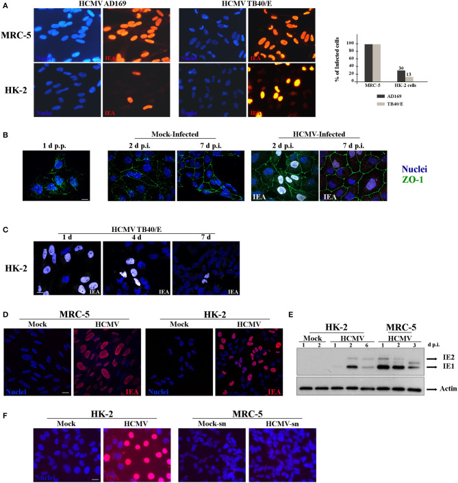
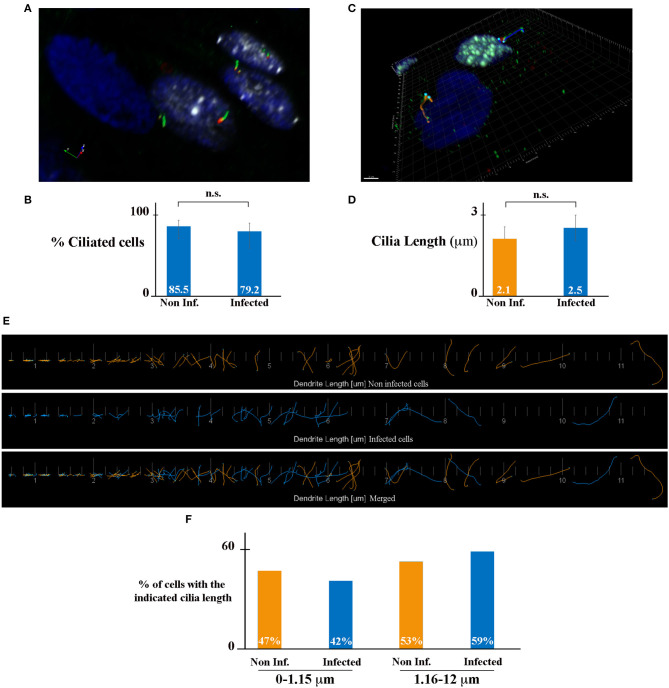
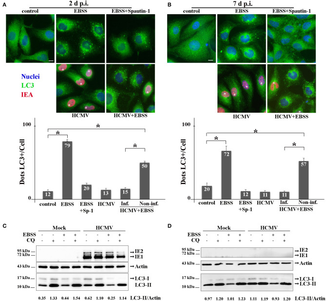
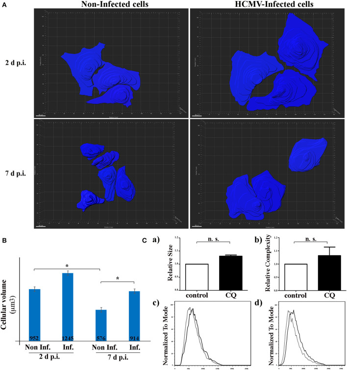
Human Cytomegalovirus (HCMV) is a frequent opportunistic pathogen in immunosuppressed patients, which can be involved in kidney allograft dysfunction and rejection. In order to study the pathophysiology of HCMV renal diseases, we concentrated on the impact of HCMV infection on human renal tubular epithelial HK-2 cells. Our aim was to develop a model of infection of HK-2 cells by using the viral strain TB40/E, that contains the extended cell tropism of clinical isolates and the efficient viral multiplication in cell culture of laboratory-adapted strains. We observed that HK-2 cells can be infected by HCMV and expressed viral antigens, but they do not produce extracellular viral particles. We then studied the interplay of HCMV with ciliogenesis and autophagy. Primary cilium (PC) is a stress sensor important to maintain renal tissue homeostasis that projects from the apical side into the lumen of tubule cells. PC formation and length were not modified by HCMV infection. Autophagy, another stress response process critically required for normal kidney functions, was inhibited by HCMV in HK-2 cells with a reduction in the autophagic flux. HCMV classically induces an enlargement of infected cells in vivo and in vitro, and we observed that HCMV infection led to an enlargement of the HK-2 cell volume. Our results constitute therefore an excellent starting point to further explore the role of these mechanisms in renal cells dysfunction.

Keywords: autophagy; cellular size; cytomegaly; cytopathic effect; human cytomegalovirus; polarization; primary cilium; renal cells.


Figures

Figure 1
Figure 1
Characterization of infection of HK-2cells by HCMV. (A) Representative images of MRC-5 and HK-2 cells infected with HCMV at MOI 1 during 48 h and immunostained for IEA (immediate-early viral antigens, in red). Two different strains, AD169 and TB40/E, were tested. Nuclei were subsequently stained with DAPI. (B) Representative images of HK-2 cells infected with HCMV or mock-infected for 2 or 7 days, and immunostained for IEA (in white) and ZO-1 (in green) to visualize cell polarization. Lack of polarization of HK-2 cells 24 h post plating (p.p.) is depicted (left panel). (C) Representative images of HK-2 cells infected with HCMV at MOI 1 at different times p.p. (1 to 7 days) and immunostained for IEA (white) and the nucleus. (D) Confocal images of MRC-5 and HK-2 cells infected with HCMV for 24 h and immunostained for IEA (red) and the nucleus. (E) Immunoblot analysis of IE1 and IE2 after HCMV infection at MOI 1 of MRC-5 or HK-2 cell lines at 1, 2, 3, or 6 days post infection (d p.i.). Actin was used as a loading control. (F) Representative images of HK-2 cells infected with HCMV for 7 days and immunostained for IEA (left panels). The non-diluted supernatants from infected (HCMV-sn) or from mock-infected HK-2 cells were tested on MRC5 cells in order to detect viral production by IEA expression (right panels). Scale bars = 10 μm. Three independent experiments were performed and statistical analysis was achieved as explained in the materials and methods section.
Figure 2
Figure 2
HCMV does not affect ciliogenesis of HK-2 cells. (A) SIM image of HCMV-infected polarized HK-2 cells immunostained for IEA (white), acetylated tubulin (PC axoneme marker, green), and γ-tubulin (PC basal body marker, red). Nuclei were subsequently stained with DAPI. (B) Percentage of ciliated cells of non-infected and infected HK-2 cells. (C) Representative 3D image of ciliated non-infected (blue nucleus) and HCMV-infected (white nucleus) HK-2 cells. The length of PC of each cell was visualized with acetylated tubulin and γ-tubulin and measured with Imaris Software. Scale bar represents 3 μm. (D) Quantification of cilia length in non-infected and infected HK-2 cells. PC length of 150 cells per condition was measured. n.s, non-significant (Student's t-test). (E) Visualization of every PC from non-infected (orange) or infected (blue) cells (separately and merged) listed in increasing order. (F) PCs were separated based on their length in two categories (0–1.15 μm) and (1.16–12 μm). Percentage of PC in these two categories in non-infected and infected cells. Three independent experiments were performed and statistical analysis was achieved as explained in the materials and methods section. n.s, non-significant (Student's t-test).
Figure 3
Figure 3
HCMV inhibits the autophagic process. (A,B) Representative images of HK-2 cells treated or not (control) with EBSS (to induce autophagy) or treated with EBSS + Spautin 1 (to block starvation-induced autophagy). HK-2 cells were also infected with HCMV at MOI 1 (and treated with EBSS for the 4 last h of infection or not). Cells were fixed after 2 days (A) or 7 days (B) and immunostained for LC3 (autophagosome marker, green) and IEA (viral protein, red). Nuclei were subsequently stained with DAPI. Scale bars represent 10 μm. Infected cells are marked with *. Quantification of LC3-positive dots per cell in each condition (lower panels). In HCMV + EBSS condition, cells expressing IEA (inf.) and neighboring non-infected cells (non-inf.) were analyzed separately. *p < 0.05 (ANOVA, with a Tukey contrast). (C,D) Immunoblot analysis of IEA and LC3-I and II in mock- or HCMV-infected HK-2 cells (2 and 7 days p.i.). Cells were treated when indicated for 4 h with EBSS and/or chloroquine (CQ). Actin was used as a loading control. A representative Western blot is depicted and LC3-II/Actin ratio is shown below the blot.
Figure 4
Figure 4
HCMV controls the size of HK-2 cells during infection. (A) 3D reconstruction of non-infected- and HCMV-infected HK-2 cells, 48 h and 7 d p.i., using over-saturated LC3 staining images to observe the complete cell and Imaris Software to represent cell size. Scale bar represent 10 μm (B) Bar graph showing quantification of the mean cell volume of non-infected and infected cells. *p < 0.05 (ANOVA, with a Tukey contrast) (C) Human epithelial HeLa cells were treated for 4 h with CQ or left in control condition. Bar graphs indicating (a) size or (b) cellular complexity of cells treated with CQ or untreated (control). Representative flow cytometry histograms showing the size (c) or cellular complexity (d) of HeLa cells treated with CQ or untreated (control). Images are representative of three independent experiments.

References

    1. Boeckh M. (2011). Complications, diagnosis, management, and prevention of CMV infections: current and future. Hematology 2011, 305–309. 10.1182/asheducation-2011.1.305 - DOI - PubMed
    1. Carney E. F. (2013). The impact of donor CMV infection on kidney graft outcome. Nat. Rev. Nephrol. 9, 434–434. 10.1038/nrneph.2013.123 - DOI - PubMed
    1. Chang T. K., Shravage B. V., Hayes S. D., Powers C. M., Simin R. T., Wade Harper J., et al. . (2013). Uba1 functions in Atg7- and Atg3-independent autophagy. Nat. Cell Biol. 15, 1067–1078. 10.1038/ncb2804 - DOI - PMC - PubMed
    1. Chaumorcel M., Lussignol M., Mouna L., Cavignac Y., Fahie K., Cotte-Laffitte J., et al. . (2012). The human cytomegalovirus protein TRS1 inhibits autophagy via its interaction with beclin 1. J. Virol. 86, 2571–2584. 10.1128/JVI.05746-11 - DOI - PMC - PubMed
    1. Chaumorcel M., Souquère S., Pierron G., Codogno P., Esclatine A. (2008). Human cytomegalovirus controls a new autophagy-dependent cellular antiviral defense mechanism. Autophagy 4, 46–53. 10.4161/auto.5184 - DOI - PubMed

Publication types