Review
. 2020 Sep 17:8:587658.
doi: 10.3389/fbioe.2020.587658. eCollection 2020.

Recent Advances of Chitosan-Based Injectable Hydrogels for Bone and Dental Tissue Regeneration

Affiliations
Review

Recent Advances of Chitosan-Based Injectable Hydrogels for Bone and Dental Tissue Regeneration

Guoke Tang et al. Front Bioeng Biotechnol. .

Abstract

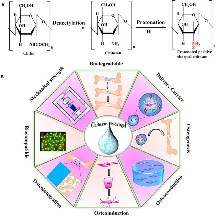
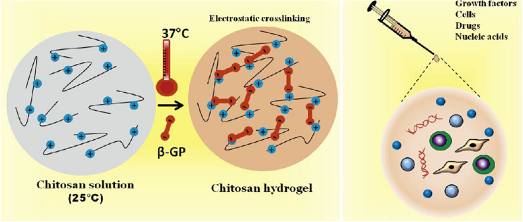
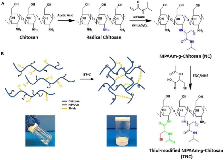
Traditional strategies of bone repair include autografts, allografts and surgical reconstructions, but they may bring about potential hazard of donor site morbidity, rejection, risk of disease transmission and repetitive surgery. Bone tissue engineering (BTE) is a multidisciplinary field that offers promising substitutes in biopharmaceutical applications, and chitosan (CS)-based bone reconstructions can be a potential candidate in regenerative tissue fields owing to its low immunogenicity, biodegradability, bioresorbable features, low-cost and economic nature. Formulations of CS-based injectable hydrogels with thermo/pH-response are advantageous in terms of their high-water imbibing capability, minimal invasiveness, porous networks, and ability to mold perfectly into an irregular defect. Additionally, CS combined with other naturally-derived or synthetic polymers and bioactive agents has proven to be an effective alternative to autologous bone and dental grafts. In this review, we will highlight the current progress in the development of preparation methods, physicochemical properties and applications of CS-based injectable hydrogels and their perspectives in bone and dental regeneration. We believe this review is intended as starting point and inspiration for future research effort to develop the next generation of tissue-engineering scaffold materials.

Keywords: bone repair; chitosan; dental tissue regeneration; injectable hydrogel; responsiveness.


Figures

FIGURE 1
FIGURE 1
(A) The chemical structures for the chitin, noncharged form of chitosan and protonated positive-charged chitosan. (B) Schematic representation of physio-chemical and biological properties of CS-based hydrogel. Reproduced from Lavanya et al. (2020) with permission from Copyright 2020 Elsevier.
FIGURE 2
FIGURE 2
Schematic representation of injectable hydrogels for treating bone loss at defective sites. Reproduced from Saravanan et al. (2019) with permission from Copyright 2019 Elsevier.
FIGURE 3
FIGURE 3
Schematic representation of pH-responsive CS-based hydrogels for bone tissue engineering. Reproduced from Zhao et al. (2019) with permission from Copyright 2019 American Chemical Society.
FIGURE 4
FIGURE 4
Schematic representation of thermal-responsive CS/β-GP hydrogels encapsulating with various growth factors, cells, drugs and nucleic acids for bone tissue engineering. Reproduced from Saravanan et al. (2019) with permission from Copyright 2019 Elsevier.
FIGURE 5
FIGURE 5
(A) Synthetic preparation of NIPAAm-g-CS. (B) Gelation mechanism of physical cross-linking (helix-coil structure) and chemical cross-linking (disulfide bonds). Reproduced from Wu et al. (2018) with permission from Copyright 2018 Elsevier.
FIGURE 6
FIGURE 6
Schematic preparation of CaCO3/MgO/CMC/BMP2 scaffolds and their applications in vivo. Reproduced from Huang et al. (2020) with permission from Copyright 2020 Elsevier.
FIGURE 7
FIGURE 7
Schematic illustration of the preparation and application of the CS/β-GP/gelatin hydrogels. Reproduced from Xu et al. (2019) with permission from Copyright 2019 Elsevier.
FIGURE 8
FIGURE 8
Effect of chitosan/bFGF scaffolds on neural differentiation of DPSCs. Immunofluorescence staining of GFAP, S100β and β-tubulin III. Reproduced from Zheng et al. (2020) with permission from Copyright 2020 Informa Healthcare.

References

    1. Abdul Rahman N., Nickles K., Gallenbach K., Dannewitz B., Ramich T., Scharf S., et al. (2019). Five-year stability of clinical attachment after regenerative treatment of infrabony defects compared to controls. J. Clin. Periodontol. 46 650–658. - PubMed
    1. Akilbekova D., Shaimerdenova M., Adilov S., Berillo D. (2018). Biocompatible scaffolds based on natural polymers for regenerative medicine. Int. J. Biol. Macromol. 114 324–333. 10.1016/j.ijbiomac.2018.03.116 - DOI - PubMed
    1. Amin S., Achenbach S. J., Atkinson E. J., Khosla S., Melton I. I. I. L. J. (2014). Trends in fracture incidence: a population-based study over 20 years. J. Bone Miner. Res. 29:581. 10.1002/jbmr.2072 - DOI - PMC - PubMed
    1. Amini A. R., Laurencin C. T., Nukavarapu S. P. (2012). Bone tissue engineering: recent advances and challenges. Crit. Rev. Biomed. Eng. 40 363–408. 10.1615/critrevbiomedeng.v40.i5.10 - DOI - PMC - PubMed
    1. Argüelles-monal W. M., Lizardi-mendoza J., Daniel F. Q., Montiel-herrera M. (2018). Chitosan derivatives: introducing new functionalities with a controlled molecular architecture for innovative materials. Polymers 10:342. 10.3390/polym10030342 - DOI - PMC - PubMed

LinkOut - more resources