. 2020 Sep 22;5(39):25440-25455.
doi: 10.1021/acsomega.0c03990. eCollection 2020 Oct 6.

Mammalian Expression and In Situ Biotinylation of Extracellular Protein Targets for Directed Evolution

Affiliations

Mammalian Expression and In Situ Biotinylation of Extracellular Protein Targets for Directed Evolution

Brian J Grindel et al. ACS Omega. .

Abstract

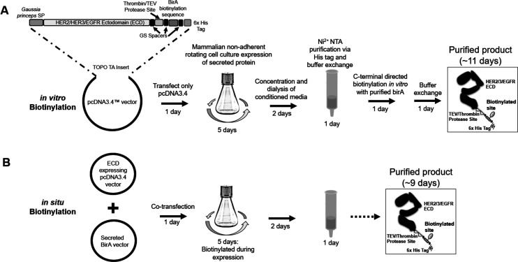
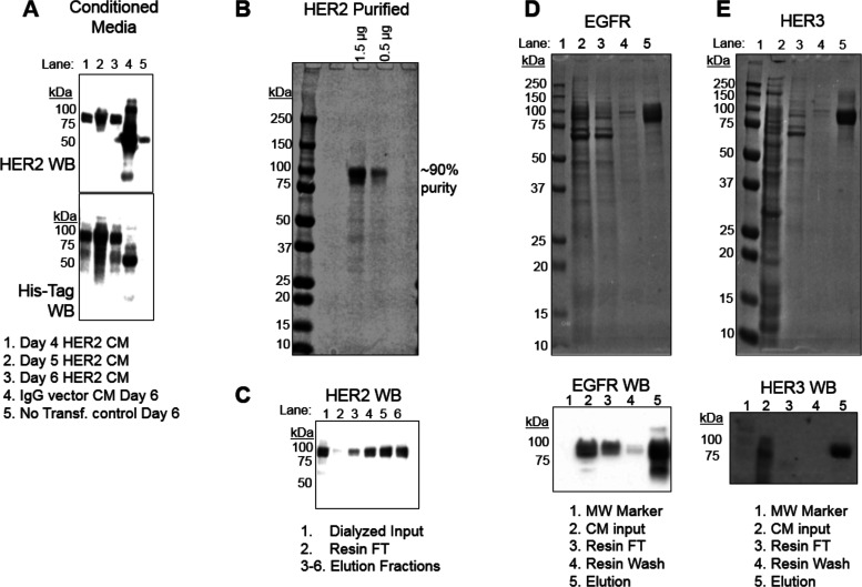
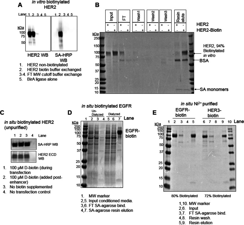
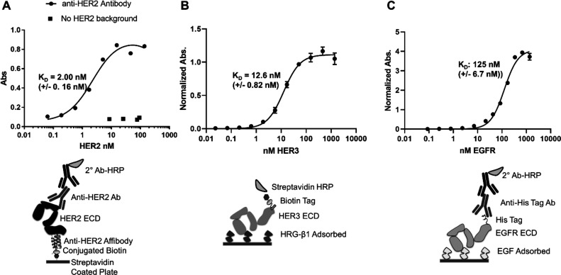
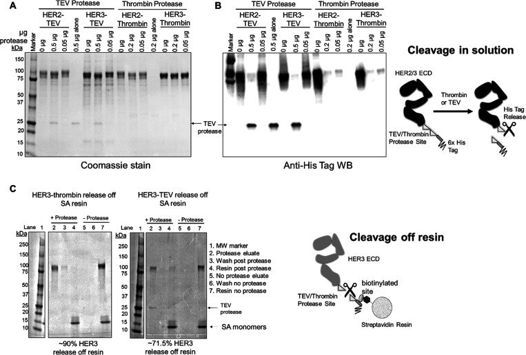
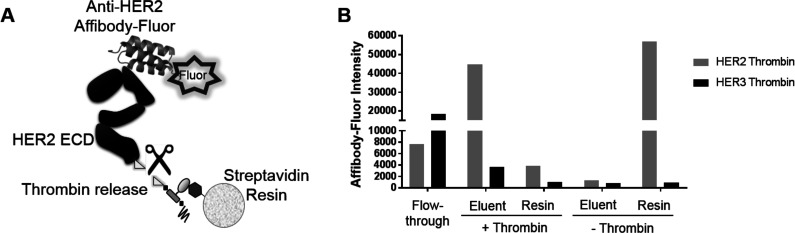
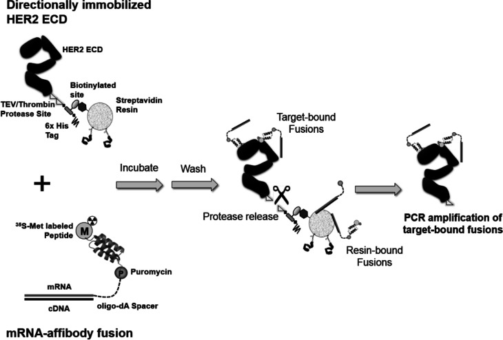
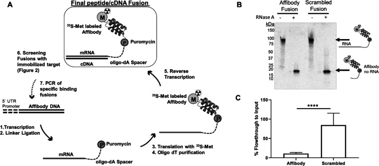
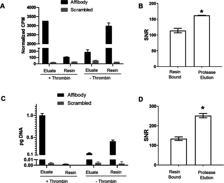
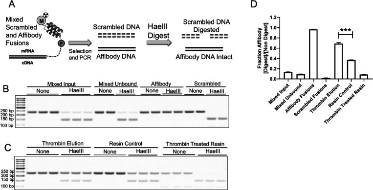
Directed evolution is a powerful tool for the selection of functional ligands from molecular libraries. Extracellular domains (ECDs) of cell surface receptors are common selection targets for therapeutic and imaging agent development. Unfortunately, these proteins are often post-translationally modified and are therefore unsuitable for expression in bacterial systems. Directional immobilization of these targets is further hampered by the absence of biorthogonal groups for site-specific chemical conjugation. We have developed a nonadherent mammalian expression system for rapid, high-yield expression of biotinylated ECDs. ECDs from EGFR, HER2, and HER3 were site-specifically biotinylated in situ and recovered from the cell culture supernatant with yields of up to 10 mg/L at >90% purity. Biotinylated ECDs also contained a protease cleavage site for rapid and selective release of the ECD after immobilization on avidin/streptavidin resins and library binding. A model mRNA display selection round was carried out against the HER2 ECD with the HER2 affibody expressed as an mRNA-protein fusion. HER2 affibody-mRNA fusions were selectively released by thrombin and quantitative PCR revealed substantial improvements in the enrichment of functional affibody-mRNA fusions relative to direct PCR amplification of the resin-bound target. This methodology allows rapid purification of high-quality targets for directed evolution and selective elution of functional sequences at the conclusion of each selection round.


Conflict of interest statement

The authors declare no competing financial interest.

Figures

Figure 1
Figure 1
Expression, purification, and biotinylation of extracellular protein targets for directed evolution. (A) Using in vitro biotinylation, the extracellular/ectodomain (ECD) protein is expressed, purified, and biotinylated by the BirA enzyme in vitro. (B) To perform in situ biotinylation, the target-encoding and BirA vectors are cotransfected resulting in target biotinylation during expression and secretion. SP is signal peptide. GS is glycine-serine repeat spacers.
Figure 2
Figure 2
EGFR family extracellular domain expression and purification in 293-F cells. (A) Time-course western blot (WB, anti-HER2 or anti-His-tag) of conditioned media (CM) indicating that the HER2 protein is expressed and secreted at high levels by day 5 post-transfection. The secreted IgG control vector is detected by the secondary HRP antibody. (B) SDS-PAGE and Coomassie staining of purified HER2 product following Ni2+-NTA purification. (C) WB of fractions from Ni2+-NTA purification. SDS-PAGE, Coomassie staining, and WB analysis of (D) EGFR and (E) HER3 following Ni2+-NTA purification. Lane descriptions are below each panel. FT is flow-through.
Figure 3
Figure 3
EGFR family receptor extracellular domains can be biotinylated both in vitro and in situ from conditioned media (CM). (A) HER2 and streptavidin (SA) horse radish peroxidase (HRP) western blot (WB) analysis of purified HER2 after in vitro biotinylation by BirA ligase. (B) SDS-PAGE and Coomassie staining of in vitro biotinylated HER2 or HER2 alone after binding to an SA-agarose resin. The first and last lanes are molecular weight (MW) standards. FT is flow-through/supernatant. (C) WB of CM at day 5 following BirA/HER2 vector cotransfection with or without supplemented d-biotin. This indicates that d-biotin supplementation is unnecessary for efficient in situ biotinylation. (D) SDS-PAGE and Coomassie staining of undialyzed (lane 2–4) or dialyzed (lane 5–7) CM from cotransfected (BirA/EGFR) 293-F cells processed through SA-agarose. This confirms that biotin must be dialyzed from the CM to achieve high-efficiency binding of the in situ biotinylated protein to SA-agarose. (E) In situ biotinylation efficiency of Ni2+-NTA-purified EGFR (lane 2–5) and HER3 (lane 6–9), as determined by SA-agarose binding.
Figure 4
Figure 4
Expressed HER2, HER3, and EGFR ECDs bind their respective ligands, demonstrating correct folding of the ECDs. (A) Expressed HER2 ECD binds to streptavidin-immobilized HER2 affibody with a KD = 2.00 nM (+/– 0.55 nM; 1.59 Hill slope), indicating that the ectodomain is properly folded. (B) Biotinylated HER3 ECD binds to immobilized heregulin β1 (HRG-β1) with a KD = 12.6 nM (+/– 0.82 nM; 1.3 Hill slope). (C) Expressed EGFR ECD binds to immobilized EGF with a KD = 125 nM (+/– 6.7 nM; 1.54 Hill slope). Binding at each concentration was measured in triplicate and the binding constants and standard deviations calculated in GraphPad Prism 8 using the specific binding with the Hill slope model. Below each titration curve is a schematic of the ELISA-based binding experiment.
Figure 5
Figure 5
Proteolytic cleavage of ECDs and release from streptavidin-agarose. Coomassie-stained SDS-PAGE (A) and anti-His-tag western blot (WB) (B) of HER2/3-TEV and HER2/3-thrombin with and without their respective proteolytic enzymes. The amount of enzyme is indicated above each lane. To the right is a schematic of the ECD and the cleavage products following proteolysis for (A) and (B). (C) Biotinylated HER3 with a thrombin/TEV cleavage site was bound to streptavidin (SA) resin and incubated with or without thrombin/TEV. Densitometry indicates 90% release by thrombin and 71.5% release by TEV. To the right is a schematic of the proteolytic release from the SA resin. These data confirm that thrombin and TEV can release the ECD from the resin without ECD proteolysis.
Figure 6
Figure 6
Selective proteolytic release of fluorescent anti-HER2 affibody from immobilized HER2 ECD. (A) Schematic of proteolytic release of HER2 with bound fluorescent affibody. (B) Fluorescent intensity of anti-HER2 affibody-Fluor solution after incubation with either SA-immobilized HER2-thrombin-biotin or HER3-thrombin-biotin followed by thrombin protease treatment (eluent). The affibody binds poorly to HER3 and therefore appears in the initial flow-through. Affibody-Fluor, in the absence of protease, stays bound to the immobilized HER2.
Figure 7
Figure 7
Directed evolution selection screen for increased target binding specificity. Site-specifically biotinylated HER2 ECD is immobilized on streptavidin resin and incubated with radiolabeled mRNA-affibody fusions. Following washing, a protease (TEV or thrombin) releases the protein target along with target-bound fusion molecules. Nonspecific or unfused (no peptide) fusions remain bound to the resin allowing for selective enrichment of HER2-binding fusions by PCR amplification.
Figure 8
Figure 8
mRNA display of anti-HER2 affibody and binding to immobilized HER2 ECD. (A) Schematic of mRNA display generation of the final 35S-labeled mRNA–affibody fusion particle. DNA encoding the anti-HER2 affibody is transcribed into mRNA and ligated to a poly-dA oligo bearing puromycin at the 3′ end. This template is translated in rabbit reticulocyte lysate resulting in the formation of an affibody–mRNA fusion. The 35S radiolabel is incorporated into the protein via [35S]-methionine in the translation mixture. Following purification with oligo dT-resin, the affibody–mRNA fusion is reverse-transcribed and panned against the immobilized HER2 ECD. The mRNA templates contain a 5′ untranslated region used to initiate translation. (B) Composition of the affibody–mRNA fusion was confirmed by SDS-PAGE autoradiography. HER2 affibody–mRNA and scrambled HER2 affibody–mRNA fusions were translated and purified as described above and incubated with RNAse A to degrade the mRNA portion. Separation by SDS-PAGE followed by autoradiography shows the correct mass for the affibody–mRNA fusion as well as the affibody-poly-dA degradation product. (C) HER2 affibody and scrambled affibody fusions were panned against immobilized HER2 ECD followed by scintillation counting of the flow-through. CPM values were converted to a percent of input for respective fusions. The scrambled fusions appeared in the flow-through, while the affibody fusions were retained on the resin and the difference between the two was found to be statistically significant (p value < .0001, seven individual experiments). This indicates that the HER2 affibody was properly folded in the context of the HER2 affibody–mRNA fusion.
Figure 9
Figure 9
HER2 affibody–mRNA fusion binding and proteolytic release on streptavidin-immobilized HER2 ECD. (A) Affibody–mRNA fusions were bound to immobilized HER2 ECD and released with thrombin. Binding and release were measured by scintillation counting of 35S-methionine activity. The HER2 affibody fusion binds HER2 and is released by the protease but remains on the bead in the absence of thrombin. The scrambled affibody–mRNA fusion does not bind to the immobilized HER2 ECD. (B) SNR is calculated by dividing the recovered activity for HER2 affibody–mRNA fusions amount by the recovered activity of the control scrambled–mRNA fusions. Eluting with thrombin results in a higher SNR compared to the direct retrieval of the fusions from the bound resin. (C) qPCR was used to analyze screening fractions that determine the mass (pg) of recovered cDNA. Eluting with thrombin releases the largest amount of affibody–mRNA fusion with limited scrambled affibody fusion release. (D) SNR values for DNA amplification of fusion products indicate higher selective recovery of genetic material via protease elution compared to direct amplification from the target-bound resin. All experimental conditions were carried out in duplicate. *; p < 0.05 (Student’s t test).
Figure 10
Figure 10
Affibody–mRNA fusions are enriched using the thrombin elution technique in a model selection. (A) Fusions were mixed 100:1 (scrambled/affibody), selected for binding to HER2, recovered directly off the resin or with thrombin, and PCR-amplified. The PCR product is then incubated with or without the HaeIII restriction enzyme. The scrambled DNA is digested by HaeIII but the affibody DNA remains intact. (B) Agarose gel of PCR product digests of the mixed input, the unbound mix flow-through, and the pure affibody and scrambled fusions. The affibody DNA remains intact after HaeIII treatment but the scrambled DNA is efficiently digested. The mixed input is mostly scrambled affibody as expected. (C) After selection, elution with thrombin yields predominantly affibody DNA (intact in HaeIII). In contrast, the thrombin-treated resin retains only scrambled DNA. The resin control also yields predominantly affibody DNA. (D) Densitometry of PCR bands was used to calculate the fraction affibody (HaeIII digest lane)/(nondigest lane), with higher values corresponding to a higher fraction of affibody DNA. While the mixed input yields mostly scrambled DNA, the thrombin elution yields mostly affibody DNA. Affibody DNA is also enriched in the resin control but significantly less so than in the thrombin elution (*** = p value < 0.001). The post-thrombin resin yields mostly scrambled DNA, indicating that the majority of the affibody–mRNA fusion has been released by thrombin activity.

Similar articles

Cited by

  • Directed Evolution of PD-L1-Targeted Affibodies by mRNA Display.
    Grindel BJ, Engel BJ, Ong JN, Srinivasamani A, Liang X, Zacharias NM, Bast RC Jr, Curran MA, Takahashi TT, Roberts RW, Millward SW. Grindel BJ, et al. ACS Chem Biol. 2022 Jun 17;17(6):1543-1555. doi: 10.1021/acschembio.2c00218. Epub 2022 May 25. ACS Chem Biol. 2022. PMID: 35611948 Free PMC article.
  • Directing evolution of novel ligands by mRNA display.
    Kamalinia G , Grindel BJ , Takahashi TT , Millward SW , Roberts RW . Kamalinia G , et al. Chem Soc Rev. 2021 Aug 21;50(16):9055-9103. doi: 10.1039/d1cs00160d. Epub 2021 Jun 24. Chem Soc Rev. 2021. PMID: 34165126 Free PMC article. Review.

References

    1. Tizei P. A. G.; Csibra E.; Torres L.; Pinheiro V. B. Selection platforms for directed evolution in synthetic biology. Biochem. Soc. Trans. 2016, 44, 1165–1175. 10.1042/BST20160076. - DOI - PMC - PubMed
    1. Goldflam M.; Ullman C. G. Recent Advances Toward the Discovery of Drug-Like Peptides De novo. Front. Chem. 2015, 3, 69. - PMC - PubMed
    1. Omidfar K.; Daneshpour M. Advances in phage display technology for drug discovery. Expert Opin. Drug Discov. 2015, 10, 651–669. 10.1517/17460441.2015.1037738. - DOI - PubMed
    1. Ståhl S.; Uhlén M. Bacterial surface display: Trends and progress. Trends Biotechnol. 1997, 15, 185–192. 10.1016/S0167-7799(97)01034-2. - DOI - PubMed
    1. Linciano S.; Pluda S.; Bacchin A.; Angelini A. Molecular evolution of peptides by yeast surface display technology. MedChemComm 2019, 10, 1569–1580. 10.1039/C9MD00252A. - DOI - PMC - PubMed