. 2020 Oct 23;23(10):101642.
doi: 10.1016/j.isci.2020.101642. Epub 2020 Oct 5.

Furin: A Potential Therapeutic Target for COVID-19

Affiliations

Furin: A Potential Therapeutic Target for COVID-19

Canrong Wu et al. iScience. .

Abstract

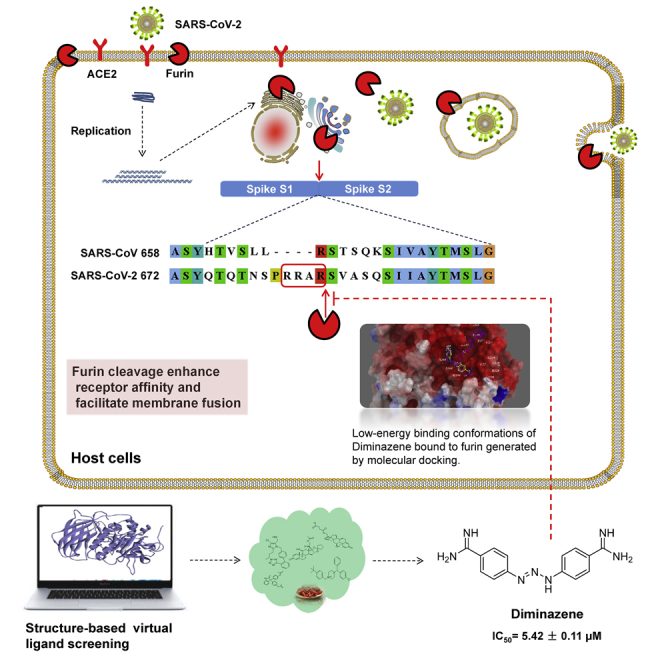
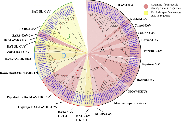
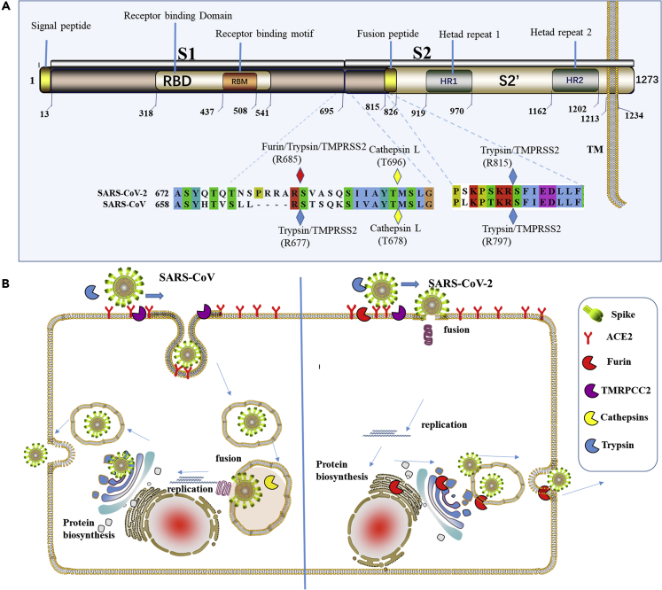
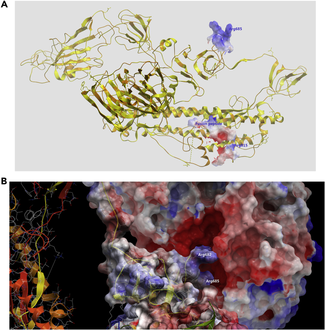
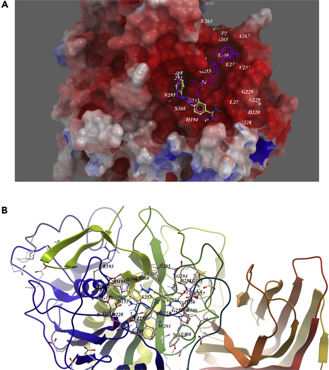
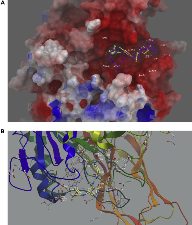
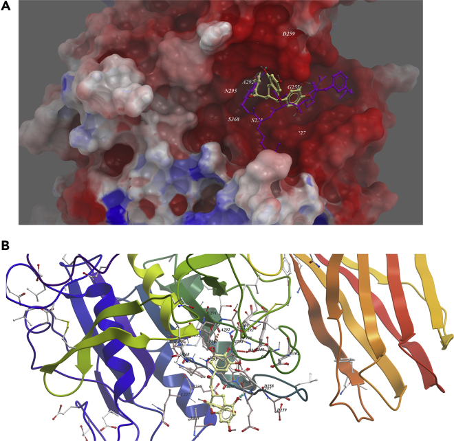
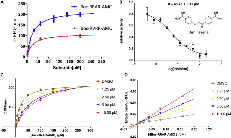
COVID-19 broke out in the end of December 2019 and is still spreading rapidly, which has been listed as an international concerning public health emergency. We found that the Spike protein of SARS-CoV-2 contains a furin cleavage site, which did not exist in any other betacoronavirus subtype B. Based on a series of analysis, we speculate that the presence of a redundant furin cut site in its Spike protein is responsible for SARS-CoV-2's stronger infectious nature than other coronaviruses, which leads to higher membrane fusion efficiency. Subsequently, a library of 4,000 compounds including approved drugs and natural products was screened against furin through structure-based virtual screening and then assayed for their inhibitory effects on furin activity. Among them, an anti-parasitic drug, diminazene, showed the highest inhibition effects on furin with an IC50 of 5.42 ± 0.11 μM, which might be used for the treatment of COVID-19.

Keywords: Computational Molecular Modelling; Disease; Drugs; Virology.


Conflict of interest statement

Authors declare no conflict of interest and no competing financial interest.

Figures

None
Graphical abstract
Figure 1
Figure 1
Evolutionary Relationships of Taxa The evolutionary history was inferred using the neighbor-joining method. The bootstrap consensus tree inferred from 500 replicates is taken to represent the evolutionary history of the taxa analyzed. Branches corresponding to partitions reproduced in less than 50% bootstrap replicates are collapsed. The evolutionary distances were computed using the Poisson correction method and in the units of the number of amino acid substitutions per site. The analysis involved 155 amino acid sequences. All positions containing gaps and missing data were eliminated. There are a total of 711 positions in the final dataset. Evolutionary analyses were conducted in MEGA7. Red shading means containing cleavage site in sequences and yellow shading means no cleavage site in sequences. All sequences are from β-coronavirus, and the four subtypes are marked in different outline colors.
Figure 2
Figure 2
Analysis of SARS-CoV-2 Spike Protein and Its Membrane Fusion Mode (A) Sequence analysis of Spike protein in SARS-CoV-2. It contains an N-terminal signal peptide, S1 and S2. S1 contains N-terminal domain and receptor-binding region. S2 is mainly responsible for membrane fusion. The C-terminal region of S2 is S2′, and it contains a fusion peptide, HR1, HR2, and a transmembrane domain. The amino acid sequence numbers of every domain are annotated below them. Cleavage sites contained in SARS-CoV and SARS-CoV-2 are marked by rhombus. (B) A schematic diagram of the process of SARS-CoV and SARS-CoV-2-infecting host cells. Those proteases are presented by sector in different colors. Furin can cleave Spike in the process of viral maturation.
Figure 3
Figure 3
Protein-Protein Docking Model of SARS-CoV-2 Spike with Furin (A) Superimposition of SARS-CoV Spike and SARS-CoV-2 Spike. Two S1/S2 protease cleavage sites and fusion peptide were shown as electrostatic surface mode. (B) Furin was docked onto the putative furin cut site (Arg685) of SARS-CoV-2 Spike. Both domains are shown as electrostatic surface mode.
Figure 4
Figure 4
Low-Energy Binding Conformations of Diminazene Bound to Furin Generated by Molecular Docking (A) Diminazene was fitted well in the active pocket of human furin, and furin was shown as electrostatic surface model. Diminazene (yellow) was overlapped with substrate analog inhibitor MI-52 (purple). (B) Detailed view of diminazene binding in the active pocket of furin.
Figure 5
Figure 5
Low-Energy Binding Conformations of Imatinib to Furin Generated by Molecular Docking (A) Imatinib was fitted well in the active pocket of human furin, and furin was shown as electrostatic surface model. Imatinib (yellow) was overlapped with substrate analog inhibitor MI-52 (purple). (B) Detailed view of Imatinib binding in the active pocket of furin.
Figure 6
Figure 6
Low-Energy Binding Conformations of ECCG to Furin Generated by Molecular Docking (A) ECCG was fitted well in the active pocket of human furin, and furin was shown as electrostatic surface model. ECCG (yellow) was overlapped with substrate analog inhibitor MI-52 (purple). (B) Detailed view of ECCG binding in the active pocket of furin.
Figure 7
Figure 7
In Vitro Enzymatic Characterization of Recombinant Human Furin Protease and Enzyme Inhibition Assay (A) Substrate saturation profiles for furin. Representative plots of the initial rate (Vi in ▵RFU/min) versus the amounts of fluorogenic substrate Boc-RVRR-AMC or Boc-RRAR-AMC. The Vmax and Km were determined by fitting the experimental data to a pseudo-first-order rate curve yielding the computed value and the best fit curve. (B) Dose response inhibition of furin activity by diminazene. (C) Substrate saturation profiles for furin at a range of concentrations of diminazene. (D) Lineweaver-Burk double-reciprocal representation of the Boc-RRAR-AMC saturation profiles for furin at a range of concentrations of Boc-RRAR-AMC.

References

    1. Belouzard S., Chu V.C., Whittaker G.R. Activation of the SARS coronavirus Spike protein via sequential proteolytic cleavage at two distinct sites. Proc. Natl. Acad. Sci. U S A. 2009;106:5871–5876. - PMC - PubMed
    1. Bosch B.J., van der Zee R., de Haan C.A., Rottier P.J. The coronavirus Spike protein is a class I virus fusion protein: structural and functional characterization of the fusion core complex. J. Virol. 2003;77:8801–8811. - PMC - PubMed
    1. Coutard B., Valle C., de Lamballerie X., Canard B., Seidah N.G., Decroly E. The Spike glycoprotein of the new coronavirus 2019-nCoV contains a furin-like cleavage site absent in CoV of the same clade. Antivir. Res. 2020;176:104742. - PMC - PubMed
    1. Dahms S.O., Arciniega M., Steinmetzer T., Huber R., Than M.E. Structure of the unliganded form of the proprotein convertase furin suggests activation by a substrate-induced mechanism. Proc. Natl. Acad. Sci. U S A. 2016;113:11196–11201. - PMC - PubMed
    1. Dahms S.O., Hardes K., Steinmetzer T., Than M.E. X-ray Structures of the proprotein convertase furin bound with substrate analogue inhibitors reveal substrate specificity determinants beyond the S4 pocket. Biochemistry. 2018;57:925–934. - PubMed

LinkOut - more resources