. 2020;2(1):115-130.
doi: 10.33597/aimm.02-1008.

Alzheimer's Disease Prevention and Treatment: Case for Optimism

Affiliations

Alzheimer's Disease Prevention and Treatment: Case for Optimism

Vladimir Volloch et al. Ann Integr Mol Med. 2020.

Abstract

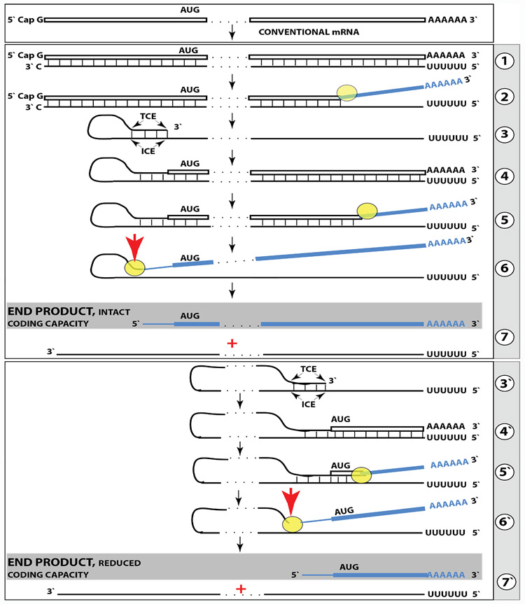
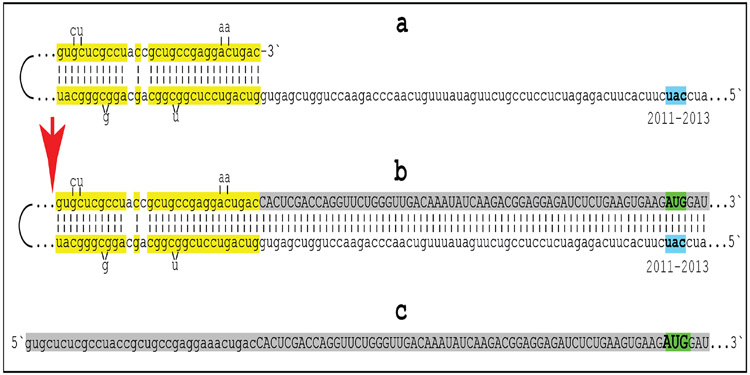
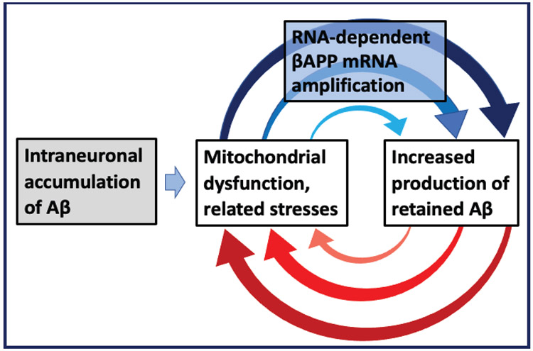
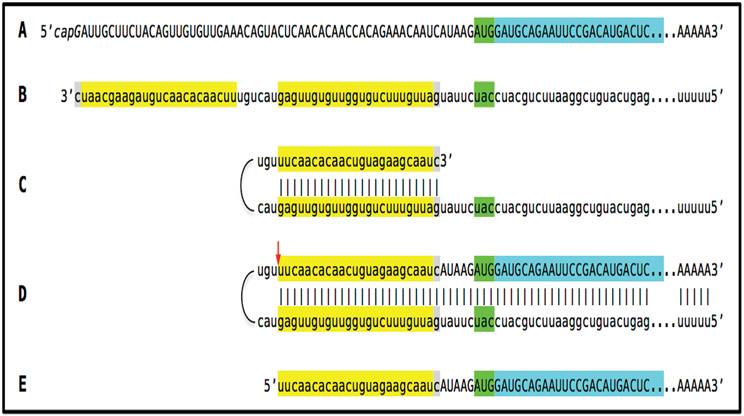
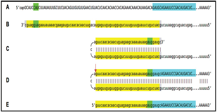
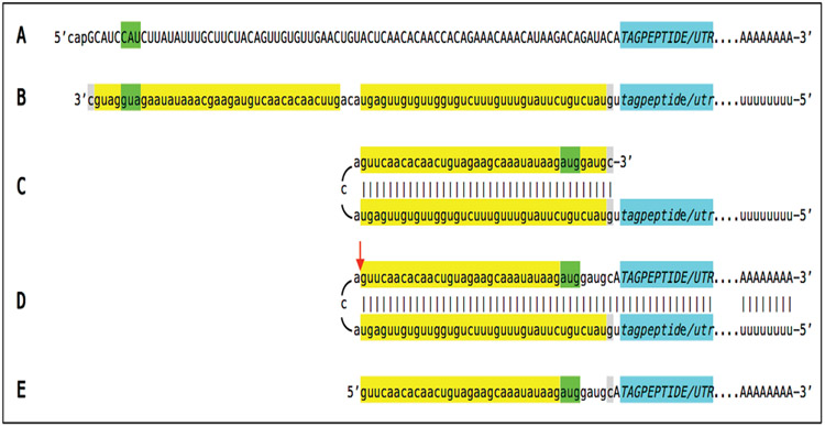
A paradigm shift is under way in the Alzheimer's field. A view of Alzheimer's disease, AD, prevailing until now, the old paradigm, maintains that it is initiated and driven by the overproduction and extracellular accumulation of beta-amyloid, Aβ; a peptide assumed to be derived, both in health and disease, solely by proteolysis of its large precursor, βAPP. In AD, according to this view, Aβ overproduction-associated neurodegeneration begins early, accumulates throughout the lifespan, and manifests symptomatically late in life. A number of drugs, designed within the framework of exceptionality of the βAPP proteolytic/secretory pathway in Aβ production in Alzheimer's disease, achieved spectacular successes in treatment, even the reversal, of AD symptoms in animal models. Without exception, they all exhibited equally spectacular failures in human clinical trials. This paradigm provides few causes for optimism with regard to prevention and treatment of AD. In its context, the disease is considered untreatable in the symptomatic phase; even prodromal cases are assumed too advanced for treatment because Aβ-triggered damages have been accumulating for preceding decades, presumably starting in the early twenties and, to be effective, this is when therapeutic intervention should commence and continue for life. The new paradigm does not dispute the seminal role of Aβ in AD but posits that beta-amyloid produced in the βAPP proteolytic/secretory pathway causes AD in humans no more than it does in non-human mammals that share this pathway with humans, accumulate Aβ as they age, but do not develop the disease. Alzheimer's disease, according to this outlook, is driven by the AD-specific pathway of Aβ production, independent of βAPP and absent in animals. Its activation, late in life, occurs through accumulation, via both cellular uptake of secreted Aβ and neuronal retention of a fraction of beta-amyloid produced in the βAPP proteolytic pathway, of intraneuronal Aβ, which triggers mitochondrial dysfunction. Cellular stresses associated with mitochondrial dysfunction, or, probably, the integrated stress response, ISR, elicited by it, activate an AD-specific Aβ production pathway. In it, every conventionally produced βAPP mRNA molecule potentially serves repeatedly as a template for production of severely 5'-truncated mRNA encoding C99 fragment of βAPP, the immediate precursor of Aβ that is processed in a non-secretory pathway, apparently in a neuron-specific manner. The resulting intraneuronally retained Aβ augments mitochondrial dysfunction, which, in turn, sustains the activity of the βAPP mRNA amplification pathway. These self-propagating Aβ overproduction/mitochondrial dysfunction mutual feedback cycles constitute the engine that drives AD and ultimately triggers neuronal death. In this paradigm, preventive treatment can be initiated any time prior to commencement of βAPP mRNA amplification. Moreover, there are good reasons to believe that with a drug blocking the amplification pathway, it would be possible not only to preempt the disease but also stop and reverse it even when early AD symptoms are already manifested. Thus, the new paradigm introduces a novel theory of Alzheimer's disease. It explains the observed discordances, determines defined therapeutic targets, provides blueprints for a new generation of conceptually distinct AD models and specifies design of a reporter for the mRNA amplification pathway. Most importantly, it offers detailed guidance and tangible hope for prevention of the disease and its treatment at the early symptomatic stages.

Keywords: AD models for the new paradigm; Asymmetric RNA-dependent βAPP mRNA amplification; Intraneuronal retention of Aβ; Reporter-based optimal AD models; Universal reporter for the mammalian RNA-dependent mRNA amplification process; βAPP-independent generation of Aβ.


Conflict of interest statement

Conflicts of Interest Authors declare no conflict of interest.

Figures

Figure 1:
Figure 1:. Projected stages of the chimeric pathway of RNA-dependent amplification of mammalian mRNA; the process can result in a 5’-truncated molecule encoding the C-terminal fragment of a conventionally encoded polypeptide.
Boxed line-sense strand RNA. Single line-antisense strand RNA. “AUG”-functional translation initiation codon (could be other than AUG). “TCE”– 3’-terminal complementary element; “ICE”– internal complementary element, both on the antisense RNA strand. Yellow circle – helicase/modifying activity complex. Blue lines (both single and boxed) – RNA strand modified and separated from its complement by a helicase complex. Red arrow – position of cleavage of the chimeric intermediate. Step 1: Synthesis of antisense strand; step 2: Strand separation; step 3: Folding of antisense strand into self-priming configuration; step 4: Extension of self-primed antisense RNA; step 5: Strand separation; step 6: Cleavage of the chimeric intermediate; step 7: End-products of RNA amplification. Steps 3’-7’ correspond to steps 3-7. Top panel: Conventional, genome-transcribed mRNA molecule. Middle panel: Projected stages of RNA-dependent mRNA amplification. The “ICE” is located within a segment of antisense RNA corresponding to the 5’UTR of conventional mRNA; the chimeric RNA end product contains the entire coding content of conventional mRNA. Bottom panel: the “ICE” is located within a segment of antisense RNA corresponding to the coding region of conventional mRNA. The amplified chimeric end product contains a 5’-truncated coding region of conventional mRNA. The translational outcome is decided by position of the first functional translation initiation codon; if in-frame, a CTF of conventional polypeptide is produced.
Figure 2:
Figure 2:. Projected topology of RNA-dependent generation of 5’-truncated mRNA encoding the C99 fragment of human beta-amyloid precursor protein.
Lowercase letters -- nucleotide sequence of the antisense RNA. Uppercase letters -- nucleotide sequence of the sense RNA. Double-stranded portions highlighted in yellow: The TCE (top) and the ICE (bottom) elements of the antisense RNA. Note that the TCE and ICE are separated by about 2000 nucleotides. “2011-2013”: nucleotide positions on the antisense RNA (starting from the complement of the AUG encoding Met1 of the βAPP) of the “uac” (highlighted in blue) corresponding to the “AUG” (highlighted in green) encoding Met671 in the βAPP mRNA. a: TCE/ICE-guided folding of the antisense βAPP RNA. 3’-terminal “c” corresponds to one of multiple transcription start sites of βAPP mRNA located 149 nucleotides upstream from its AUG initiation codon [28]; note that such folding configuration would accommodate the additional 3’-terminal “C”, a transcript of the capG of βAPP mRNA (not shown). b: Extension of self-primed antisense RNA into sense RNA and cleavage (red arrow; may also occur at one of the TCE/ICE mismatches), after strand separation, of the chimeric intermediate. c: Chimeric RNA end product contains 5’terminal antisense segment extending into severely 5’-truncated βAPP mRNA. Its translation initiates from the “AUG” (highlighted in green and encoding Met671 in conventional βAPP mRNA) immediately preceding the beta amyloid-encoding region.
Figure 3:
Figure 3:. The engine that drives AD: Self-propagating mutual feedback cycles of mitochondrial dysfunction-mediated overproduction of Aβ and vice versa in Alzheimer’s disease.
Highlighted in grey: Intraneuronal accumulation of Aβ via both cellular uptake of secreted peptide and retention of a fraction of beta-amyloid produced in the βAPP proteolytic pathway; Highlighted in blue: Asymmetric RNA-dependent βAPP mRNA amplification, a molecular basis of Aβ overproduction in Alzheimer’s disease. Horizontal arrow: Intraneuronal accumulation of Aβ critical levels acts as a starter motor that initiates the self-sustainable engine that drives AD. Arched arrows: Self-propagating mutual feedback cycles; Red arches: Intraneuronal Aβ-mediated induction of mitochondrial dysfunction and related stresses; Blue arches: Mitochondrial dysfunction/related stresses-mediated RNA-dependent amplification of βAPP mRNA resulting in overproduction of intraneuronally retained Aβ.
Figure 4:
Figure 4:. Dynamics of Alzheimer’s disease in two paradigms.
Left panels: Dynamics of Aβ production; Right panels: Dynamics of neurodegeneration. Blue lines: Levels of beta-amyloid; Red lines: Extent of neurodegeneration; Black lines: Indicator lines, no significant neurodegeneration; Red blocks: Symptomatic manifestation of AD. T: Threshold of symptomatic manifestation of AD (reflects levels of extracellular Aβ and consequent extent of neurodegeneration); T1: Threshold of activation of RNA-dependent βAPP mRNA amplification (reflects both intraneuronal Aβ levels and the consequent extent of Aβ–coupled mitochondrial dysfunction); Numerous genetic factors such as the occurrence of various alleles of ApoE gene, as well as certain epigenetic factors, influence the age when thresholds T and T1 are reached, hence, the fanning lines.T2: Threshold of symptomatic occurrence of AD (reflects levels of intraneuronal Aβ,degree of mitochondrial dysfunction and consequent extent of neurodegeneration). Panels A, B: View of the dynamics of AD in the old paradigm (A: Dynamics of SAD; B: Dynamics of FAD). Levels of extracellular Aβ increase, neurodegeneration starts early and accumulates throughout the life. When threshold T is reached, AD symptoms manifest. Panels C, D: The outlook on the dynamics of AD in the new paradigm (C: Dynamics of SAD; D: Dynamics of FAD). Levels of intraneuronal Aβ increase, the extent of mitochondrial dysfunction and related stresses reach threshold T1 and RNA-dependent βAPP mRNA amplification is activated. There is no significant neurodegeneration until after a lag period (when amplified RNA encoding the C99 fragment of βAPP accrues and intraneuronal Aβ further accumulates) following the crossing of T1 threshold and activation of βAPP mRNA amplification; when the extent of neurodegeneration reaches threshold T2, AD symptoms manifest. Panel E: Dynamics of Aβ production and neurodegeneration in non-human mammals: T1 threshold is crossed but βAPPP mRNA is not amplified because it is not eligible for RNA-dependent amplification process. There is no significant neurodegeneration; T2 threshold is not reached, no AD symptoms manifest, no disease occurs. Note: Scenario depicted in panel(E) would occur in humans not susceptible to Alzheimer’s disease due to variations in βAPP TSSs utilization or for other reasons, or when the βAPP mRNA amplification pathway of Aβ production is effectively interfered with by therapeutic intervention.
Figure 5:
Figure 5:. Example of construct-expressed RNA-dependent amplification-eligible mRNA encoding Aβ or C99 fragment of βAPP and projected stages of its amplification.
Uppercase letters: Nucleotide sequence of the sense RNA. Lowercase letters: Nucleotide sequence of the antisense RNA. Highlighted in blue: Aβ- or C99-encoding region of mRNA. Highlighted in green: 5’AUG3’ translation initiation codon in an optimal context or its complement on the opposite RNA strand. Highlighted in yellow: the 3’-terminal complementary element, TCE, and the internal complementary element, ICE, of the antisense RNA. Highlighted in gray: 3’-terminal “C” not encoded in the DNA, a transcript of the cap”G” of mRNA and matching “G” at the 5’ end of the ICE on the antisense RNA. Red arrow: Position of cleavage of chimeric intermediate following strand separation. A: mRNA encoding Aβ or C99 fragment of βAPP. B: Antisense RNA; note that it contains an additional 3’-terminal “C” not encoded in the genome, a transcript of the cap”G” of mRNA. C: Antisense RNA folded into self-priming configuration; note that the additional 3’-terminal “C” is accommodated by a “G” at the 5’end of the ICE. D: Extension of self-primed antisense RNA generating chimeric intermediate containing covalently bound sense and antisense RNA strands; position of cleavage following strand separation is indicated by red arrow. E: Chimeric RNA end product of RNA-dependent mRNA amplification; it consists of an antisense segment, the TCE, continued into a sense-orientation sequence containing the rest of the 5’UTR and the Aβ- or C99-coding sequence preceded by the AUG translation initiation codon and followed by the 3’UTR and 3’- poly(A).
Figure 6:
Figure 6:. Example of “silent” RNA transcript from a construct producing Aβ or C99 fragment solely in the RNA-dependent amplification pathway; projected amplification stages resulting in functional chimeric mRNA encoding Aβ or C99.
Uppercase letters: Nucleotide sequence of the sense RNA. Lowercase letters: Nucleotide sequence of the antisense RNA. Highlighted in blue: Aβ- or C99-encoding region of mRNA. Highlighted in green: 5’AUG3’ in an optimal translation initiation context or its complement on the opposite RNA strand. Highlighted in yellow: 3’-terminal complementary element, TCE, and internal complementary element, ICE, of the antisense RNA. Highlighted in gray: 3’-terminal “C” not encoded in the DNA, a transcript of the cap”G” of mRNA and matching “G” at the 5’ end of the ICE on the antisense RNA. Red arrow: Position of cleavage of chimeric intermediate following strand separation. A–Translationally silent RNA encoding Aβ or C99 fragment of βAPP; note that the “AUG” immediately preceding Aβ-coding in endogenous mRNA has been removed and replaced by the “ACA”, and there is no functional translation initiation codon in-frame with and upstream from Aβ-coding segment. B - Antisense RNA; note that it contains 5’aug3’ followed in the 3’ direction by sequence encoding N-terminus of Aβ at its 3’end. C - Antisense RNA folded into self-priming configuration; note that 5’aug3’ is accommodated by the 3’ugu5’, a complement of the “ACA” in the sense strand. D - Extension of self-primed antisense RNA generating chimeric intermediate containing covalently bound sense and antisense RNA strands; position of cleavage following strand separation is indicated by red arrow. E - Chimeric RNA end product of RNA-dependent mRNA amplification; note that it consists of the antisense portion encoding the “aug” translation initiation codon in optimal translation initiation context and the N-terminus of resulting polypeptide (Aβ or C99), whereas the sense RNA portion encodes the rest of a polypeptide and contains 3’UTR and 3’-terminal poly(A).
Figure 7:
Figure 7:. Example of “silent” RNA transcript from a reporter construct producing tag peptide only when the RNA-dependent mRNA amplification pathway is operational; projected stages leading to generation of functional chimeric mRNA encoding tag peptide.
Uppercase letters: Nucleotide sequence of the sense RNA. Lowercase letters: Nucleotide sequence of the antisense RNA. TAGPEPTIDE/UTR, highlighted in blue, denotes nucleotide sequence encoding a tag peptide followed by 3’UTR and lacking the AUG translation initiation codon; tagpeptide/utr, highlighted in blue, denotes antisense complement of the TAGPEPTIDE/UTR nucleotide sequence. Highlighted in green: 5’AUG3’ in an optimal translation initiation context or its complement on the opposite RNA strand. Highlighted in yellow: the 3’-terminal complementary element, TCE, and internal complementary element, ICE, of the antisense RNA. Highlighted in gray: 3’-terminal “C” not encoded in the DNA, a transcript of the cap”G” of mRNA and matching “G” at the 5’ end of the ICE on the antisense RNA. Red arrow: Position of cleavage of chimeric intermediate following strand separation. A – Translationally silent RNA transcript encoding tag peptide and lacking the AUG translation initiation codon; there is also no functional translation initiation codon upstream from and in-frame with the tag peptide-encoding segment. B - Antisense RNA; note that it contains 5’aug3’ in the optimal translation initiation context. C - Antisense RNA folded into self-priming configuration; note that 5’aug3’ is accommodated by the 3’ugu5’, a complement of the “ACA” in the sense strand. D - Extension of self-primed antisense RNA generating chimeric intermediate containing covalently bound sense and antisense RNA strands; position of cleavage following strand separation is indicated by red arrow. E - Chimeric mRNA end product of RNA-dependent mRNA amplification; note that the “aug” translation initiation codon in optimal translation initiation context provided by the antisense portion is in-frame with the tag peptide-encoding nucleotide sequence in the sense portion of the chimeric RNA end product.

Similar articles

Cited by

References

    1. Conan-Doyle A. The sign of the four. Lippincott’s Monthly Magazine. 1890.
    1. Volloch V. A mechanism for ß-amyloid overproduction in Alzheimer's disease: Precursor-independent generation of ß-amyloid via antisense RNA-primed mRNA synthesis. FEBS Lett. 1996;390:124–8. - PubMed
    1. Volloch V. Mechanism for ß-amyloid overproduction in sporadic Alzheimer’s Disease: Possible antisense RNA-mediated generation of a 5’-truncated ßAPP mRNA encoding 12 kDa C-terminal fragment of ßAPP, the immediate precursor of Aß In: Molecular Mechanisms of Dementia. 1997, Wasco W and Tanzi R, Eds.
    1. Volloch V. Possible mechanism for resistance to Alzheimer's disease (AD) in mice suggests new approach to generate a mouse model for sporadic AD and may explain familial resistance to AD in man. Exp Neurobiol. 1997;144:214–8. - PubMed
    1. Volloch V, Rits S. Results of beta-secretase-inhibitor clinical trials support amyloid precursor protein-independent generation of beta-amyloid in sporadic Alzheimer's disease. Med Sci. 2018. - PMC - PubMed

LinkOut - more resources