. 2021 Mar;31(2):239-252.
doi: 10.1111/bpa.12905. Epub 2020 Nov 2.

Cerebellar developmental deficits underlie neurodegenerative disorder spinocerebellar ataxia type 23

Affiliations

Cerebellar developmental deficits underlie neurodegenerative disorder spinocerebellar ataxia type 23

Cleo J L M Smeets et al. Brain Pathol. 2021 Mar.

Abstract

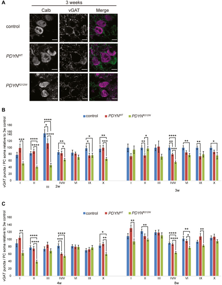
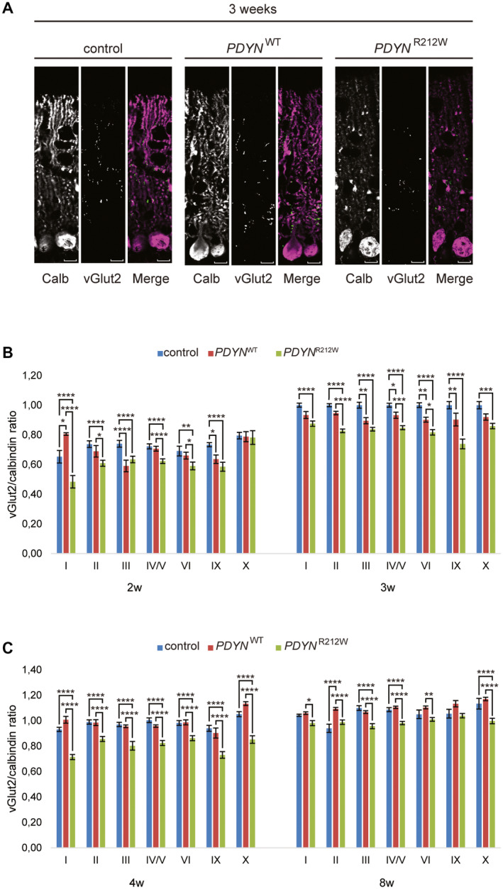
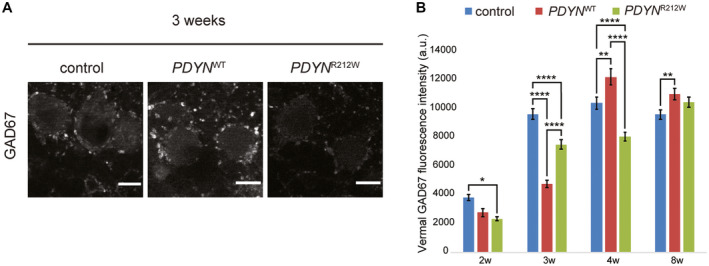
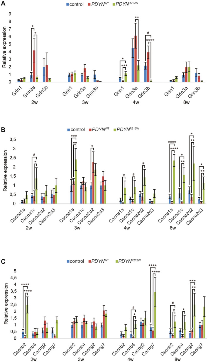
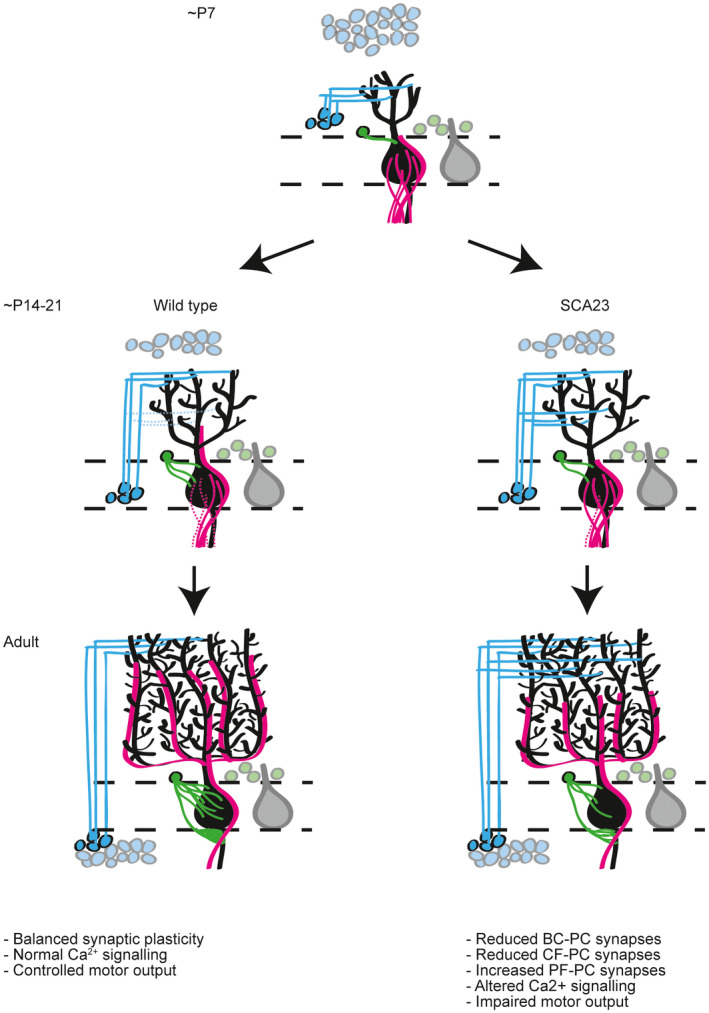
Spinocerebellar ataxia type 23 (SCA23) is a late-onset neurodegenerative disorder characterized by slowly progressive gait and limb ataxia, for which there is no therapy available. It is caused by pathogenic variants in PDYN, which encodes prodynorphin (PDYN). PDYN is processed into the opioid peptides α-neoendorphin and dynorphins (Dyn) A and B; inhibitory neurotransmitters that function in pain signaling, stress-induced responses and addiction. Variants causing SCA23 mostly affect Dyn A, leading to loss of secondary structure and increased peptide stability. PDYNR212W mice express human PDYN containing the SCA23 variant p.R212W. These mice show progressive motor deficits from 3 months of age, climbing fiber (CF) deficits from 3 months of age, and Purkinje cell (PC) loss from 12 months of age. A mouse model for SCA1 showed similar CF deficits, and a recent study found additional developmental abnormalities, namely increased GABAergic interneuron connectivity and non-cell autonomous disruption of PC function. As SCA23 mice show a similar pathology to SCA1 mice in adulthood, we hypothesized that SCA23 may also follow SCA1 pathology during development. Examining PDYNR212W cerebella during development, we uncovered developmental deficits from 2 weeks of age, namely a reduced number of GABAergic synapses on PC soma, possibly leading to the observed delay in early phase CF elimination between 2 and 3 weeks of age. Furthermore, CFs did not reach terminal height, leaving proximal PC dendrites open to be occupied by parallel fibers (PFs). The observed increase in vGlut1 protein-a marker for PF-PC synapses-indicates that PFs indeed take over CF territory and have increased connectivity with PCs. Additionally, we detected altered expression of several critical Ca2+ channel subunits, potentially contributing to altered Ca2+ transients in PDYNR212W cerebella. These findings indicate that developmental abnormalities contribute to the SCA23 pathology and uncover a developmental role for PDYN in the cerebellum.

Keywords: GABAergic transmission; ataxia; climbing fibers; development; prodynorphin.


Conflict of interest statement

The authors declare that they have no conflict of interest.

Figures

Figure 1
Figure 1
GABAergic innervation of Purkinje cells is reduced in PDYN R212W mice. GABAergic presynaptic innervation of Purkinje cell (PC) soma in the vermis was determined by vGAT and Calbindin immunostaining, respectively. (A) Representative fluorescence images of vermal lobule IV/V of control, PDYN WT and PDYN R212W mice at 3 weeks of age stained with anti‐calbindin antibody (magenta) and anti‐vGAT (green) antibody. Scale bar = 20 μm. (B‐C) Quantification of vGAT vesicles on PC somas, relative to control mice of 3 weeks of age, in vermal lobules I, II, III, IV/V, VI, IX and X of control, PDYN WT and PDYN R212W mice of 2, 3, 4 and 8 weeks of age (n = 5 cells per section per lobule [minimum 20 cells] per genotype). PDYN R212W mice show reduced amounts of somatic vGAT vesicles early in development. *P < 0.05, **P < 0.01, ***P < 0.001 and ****P < 0.0001.
Figure 2
Figure 2
Climbing fiber synapse elimination is impaired in PDYN R212W mice. The presynaptic innervation of Purkinje cells (PC) by climbing fibers (CF) in the vermis during development was assessed by Calbindin and vGlut2 immunostaining, respectively. (A) Representative fluorescence images of vermal lobule X of control, PDYN WT and PDYN R212W mice at 2 and 3 weeks of age stained with anti‐Calbindin (magenta) and anti‐vGlut2 (green) antibody. Scale bar = 20 μm (B‐D) Quantification of the vGlut2 puncta per PC soma in vermal lobules II, III, IV/V, VI, IX and X of control (B), PDYN WT (C) and PDYN R212W (D) mice at 2, 3, 4 and 8 weeks of age . Control and PDYN WT mice show a marked reduction of vGlut2 synapses over time in all lobules. In contrast, PDYN R212W mice display a delay in reduction of vGlut2 synapses over time in all lobules except lobule X, which shows no decline of vGlut2 synapses (n = 5 cells per section per lobule [minimum 20 cells] per genotype). *P < 0.05, **P < 0.01, ***P < 0.001 and ****P < 0.0001.
Figure 3
Figure 3
PDYN R212W mice show developmental climbing fiber deficits. The distribution of climbing fibers (CF) over the Purkinje cell (PC) dendritic tree in the vermis was assessed by vGlut2 and Calbindin immunostaining, respectively. (A) Representative confocal images of vermal lobule IV/V of control, PDYN WT and PDYN R212W mice at 3 weeks of age stained with anti‐Calbindin (magenta) and anti‐vGlut2 (green) antibody. (B–C) Quantification of the relative height of vGlut2 compared to Calbindin staining in vermal lobules I, II, III, IV/V, VI, IX and X of control, PDYN WT and PDYN R212W mice of 2 and 3 weeks of age (n = 4–7, per genotype). At 2 weeks of age, PDYN R212W mice show significant loss of CF‐PC synapses in all lobules except lobule X. At 3 weeks of age, lobule X also displayed reduced CF‐PC synapses in PDYN R212W mice. All lobules showed reduced CF‐PC synapses in PDYN R212W mice at 4 weeks of age, while at 8 weeks of age, significance in lobule IX was lost. *P < 0.05, **P < 0.01, ***P < 0.001 and ****P < 0.0001.
Figure 4
Figure 4
Altered vGlut1 protein levels indicate developmental abnormalities in the vermis of PDYN R212W mice. (A,B) The left panel shows representative immunoblots of control, PDYN WT and PDYN R212W vermal protein lysates, stained or vGlut1 and actin. The right panel shows quantification of these immunoblots using ImageLab software. vGlut1 protein levels were increased in PDYN R212W at 2, 3 and 4 weeks compared with control mice and at 2, 4 and 8 weeks of age compared with PDYN WT mice (n = 3 per genotype). # 0.10> p> 0.05, *P < 0.05, **P < 0.01.
Figure 5
Figure 5
PDYN R212W mice show reduced GABAergic transmission. The GABAergic inhibitory transmission by interneurons in the vermis was assessed with GAD67 immunostaining. (A) Representative fluorescence images of vermal lobule IV/V of control, PDYN WT and PDYN R212W mice at 3 weeks of age stained with anti‐GAD67 antibody. Scale bar = 20 μm (B) Quantification of the relative fluorescence intensity of GAD67 in the total vermis of control, PDYN WT and PDYN R212W mice of 2, 3, 4 and 8 weeks of age (n = 20–30 images per genotype, minimum 24 measurements). PDYN R212W vermis shows reduced GAD67 fluorescence intensity as compared to control mice at 2, 3 and 4 weeks of age. *P < 0.05, **P < 0.01 and ****P < 0.0001.
Figure 6
Figure 6
Altered expression of Ca2+ channel subunits suggests a compensatory mechanism in PDYN R212W mice. Quantification of RT‐qPCR data from 2, 3, 4 and 8‐week‐old cerebella from control, PDYN WT and PDYN R212W mice (n = 6, per genotype). (A) At 4 weeks of age, Grin1 showed elevated expression in PDYN R212W as compared to PDYN WT and control mice. Simultaneously, Grin3a and –b expression was significantly reduced in these mice as compared to control and PDYN WT mice, respectively. For Grin3a, expression was increased in PDYN WT compared with control and PDYN R212W mice, but this was likely due to expression of PDYN‐WT. (B) Cacna1a expression was elevated at 3, 4 and 8 weeks of age in PDYN R212W as compared to control and PDYN WT mice, while Cacna1c expression was increased at 2, 4 and 8 weeks of age. In 3, 4 and 8‐week‐old PDYN R212W mice, Cacna2d2 was elevated, and at 8 weeks of age, Cacna2a3 expression was elevated in PDYN R212W mice as well compared with control and PDYN WT mice. (C) Cacnb2 expression was increased at 2 and 8 weeks of age in PDYN R212W mice compared to both control and PDYN WT mice, while Cacnb4 expression was enhanced at 4 weeks of age. For Cacng2 and Cacng7, expression was elevated at 8 and 4 weeks of age, respectively. # 0.10> p> 0.05, *P < 0.05, **P < 0.01, ***P < 0.001 and ****P < 0.0001.
Figure 7
Figure 7
Cerebellar developmental abnormalities underlie SCA23. Under normal conditions, cerebellar development progresses in highly regulated, clear phases (left panels). In SCA23 cerebella, we have observed several disruptions of cerebellar development. First, basket cells (in green) form fewer somatic synapses in SCA23 cerebella. Second, climbing fibers (CFs, in pink) display a delay in CF synapse elimination, as well as a failure to reach terminal height to innervate the proximal Purkinje cell (PC) dendrites. These deficits likely lead to the third disruption, an increase in parallel fiber‐PC synapses (in blue). These developmental abnormalities, combined with our other findings, very likely lead to altered Ca2+ signaling and, consequently, impaired motor output and ataxia.

Similar articles

Cited by

References

    1. Altier C, Zamponi GW (2006) Opioid, cheating on its receptors, exacerbates pain. Nat Neurosci 9:1465–1467. - PubMed
    1. Bakalkin G, Watanabe H, Jezierska J, Depoorter C, Verschuuren‐Bemelmans C, Bazov I et al (2010) Prodynorphin mutations cause the neurodegenerative disorder spinocerebellar ataxia type 23. Am J Hum Genet 87:593–603. - PMC - PubMed
    1. Barclay J, Balaguero N, Mione M, Ackerman SL, Letts VA, Brodbeck J et al (2001) Ducky mouse phenotype of epilepsy and ataxia is associated with mutations in the Cacna2d2 gene and decreased calcium channel current in cerebellar purkinje cells. J Neurosci 21:6095–6104. - PMC - PubMed
    1. Bhat S, Dao DT, Terrillion CE, Arad M, Smith RJ, Soldatov NM, Gould TD. (2012) CACNA1C (Cav1.2) in the pathophysiology of psychiatric disease. Prog Neurobiol 99:1–14. - PMC - PubMed
    1. Brown AM, Arancillo M, Lin T, Catt DR, Zhou J, Lackey EP et al (2019) Molecular layer interneurons shape the spike activity of cerebellar Purkinje cells. Sci Rep 9:1742. - PMC - PubMed

Publication types

Supplementary concepts