Review
. 2021 Jun 11;7(6):1332-1350.
doi: 10.1021/acsinfecdis.0c00552. Epub 2020 Oct 12.

Inhibition of Influenza Virus Polymerase by Interfering with Its Protein-Protein Interactions

Affiliations
Review

Inhibition of Influenza Virus Polymerase by Interfering with Its Protein-Protein Interactions

Serena Massari et al. ACS Infect Dis. .

Abstract

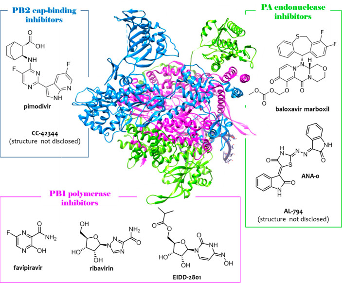
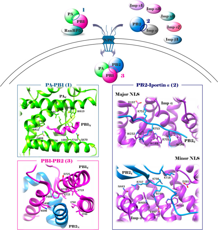
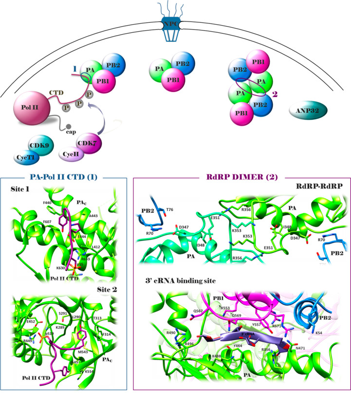
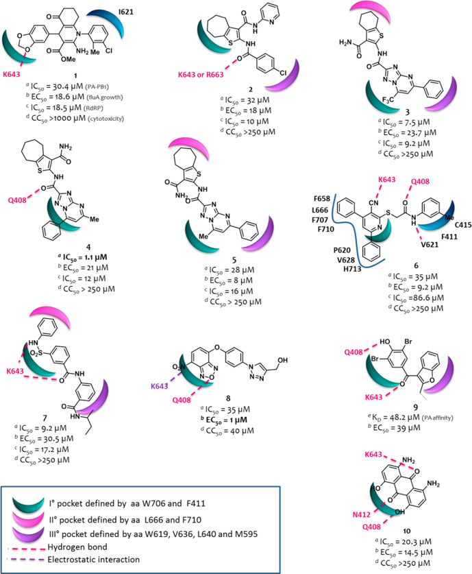
Influenza (flu) virus is a serious threat to global health with the potential to generate devastating pandemics. The availability of broad spectrum antiviral drugs is an unequaled weapon during pandemic events, especially when a vaccine is still not available. One of the most promising targets for the development of new antiflu therapeutics is the viral RNA-dependent RNA polymerase (RdRP). The assembly of the flu RdRP heterotrimeric complex from the individual polymerase acidic protein (PA), polymerase basic protein 1 (PB1), and polymerase basic protein 2 (PB2) subunits is a prerequisite for RdRP functions, such as mRNA synthesis and genome replication. In this Review, we report the known protein-protein interactions (PPIs) occurring by RdRP that could be disrupted by small molecules and analyze their benefits and drawbacks as drug targets. An overview of small molecules able to interfere with flu RdRP functions exploiting the PPI inhibition approach is described. In particular, an update on the most recent inhibitors targeting the well-consolidated RdRP PA-PB1 subunit heterodimerization is mainly reported, together with pioneer inhibitors targeting other virus-virus or virus-host interactions involving RdRP subunits. As demonstrated by the PA-PB1 interaction inhibitors discussed herein, the inhibition of flu RdRP functions by PPI disrupters clearly represents a valid means to identify compounds endowed with a broad spectrum of action and a reduced propensity to develop drug resistance, which are the main issues of antiviral drugs.

Keywords: PA−PB1; PA−Pol II CTD; PB1−PB2; PB1−RanBP5; PB2−ANP32; PB2−importin-α; RNA-dependent RNA polymerase; anti-influenza small molecules; protein−protein interface inhibitors.


Conflict of interest statement

The authors declare no competing financial interest.

Figures

Figure 1
Figure 1
Crystal structure of flu A RdRP determined from bat flu A H17N10 strain (pdb: 4WSB(30)) and chemical structures of inhibitors of PA (green), PB2 (blue), and PB1 (magenta) subunits, approved or in the pipeline. The overall RdRP is U shaped with the PAN endonuclease and PB2 cap-binding domains being the two upper protuberances, the PAC domain being the bottom, and the PB1 polymerase domain filling the interior. Among the reported compounds, PA endonuclease inhibitor baloxavir marboxil and PB1 inhibitor favipiravir have been approved. The figure is author created, and the RdRP structure has been adapted from the pdb mentioned above and drawn by using UCSF Chimera package.
Figure 2
Figure 2
Schematic representation of flu RdRP subunit nuclear localization and heterotrimerization (upper side): in the cytoplasm, PA and PB1 form a heterodimer (extensive interactions occurring between PAC and PB1N); then, the PA–PB1 heterodimer associates with RanBP5 (interaction occurring at the PB1N bipartite NLS), and PB2 associates with importin-α1, -α3, -α5, or -α7 (interaction occurring at the PB2 NLS), which then binds to importin-β1, to enter within the nucleus; finally, once in the nucleus, PA–PB1 and PB2 associate (extensive interactions occurring between PB1C and PB2N) to form the whole RdRP heterotrimer. For clarity, the RdRP is shown alone and not in the context of the vRNP. Crystal structures of PA–PB1 (pdb: 3CM8(72)), PB2–importin-α (as an example, the PB2–importin-α7 complex was shown; pdb: 4UAD(61)), and PB1–PB2 (pdb: 2ZTT(76)) interfaces (lower side). PA subunit, green; PB2 subunit, blue; PB1 subunit, magenta. The figure is author created, and the structures have been adapted from the pdb mentioned above and drawn by using the UCSF Chimera package.
Figure 3
Figure 3
Schematic representation of the flu RdRP association with Pol II during vRNA transcription and dimerization during vRNA replication (upper side): once heterotrimerization has occurred in the nucleus, RdRP performs both the processes of transcription and replication; during transcription of viral mRNA, the specific interaction between the PAC and host Pol II CTD is required to enable the process of cap-snatching; during replication, a new RdRP is synthesized and associates with the resident RdRP to form a dimer, which is required for the synthesis of cRNA from vRNA (interactions occurring between the PAC loop of the two RdRPs and PAC of one RdRP and PB2 loop of the other); finally, the association of PB2 and host factor ANP32 promote the replication of vRNA from cRNA (interaction occurring at the PB2 627 domain). For clarity, the RdRP is shown alone and not in the context of the vRNP. Crystal structures of the PA–Pol II CTD interface (pdb: 5M3H(81)), RdRP–RdRP interface (pdb: 6QPG(33)), and 3′ cRNA binding site (pdb: 6QX3(33)) (lower side). PA subunit, green; PB2 subunit, blue; PB1 subunit, magenta. The figure is author created, and the structures have been adapted from the pdb mentioned above and drawn by using the UCSF Chimera package.
Figure 4
Figure 4
Structures of representative compounds reported as PA–PB1 inhibitors until 2015. aThe IC50 value represents the compound concentration that reduces the PA–PB1 complex formation by 50% (ELISA assay); bthe EC50 value represents the compound concentration that inhibits 50% of flu A replication (PRA assay); cthe IC50 value represents the compound concentration that reduces by 50% the activity of flu A virus RNA polymerase (minireplicon assay); dthe CC50 value represents the compound concentration that inhibits 50% of cell growth (MTT assay); ethe Kd value represents the dissociation constant of the compound with the PA cavity. The predicted binding mode of all the molecules, with the exception of 6, in the PA cavity from structure 3CM8 was generated using FLAP. The predicted binding mode of 6 in the PA cavity (generated by Glide and GOLD) was reported as in the original paper. The figure is author created, while the original figures reporting the binding pose of compounds 110 are reported in Figures S1 and S2.
Figure 5
Figure 5
Structures and activities of PA–PB1 interaction inhibitors identified by Yuan et al. (in the blue box) and Watanabe et al. (in the magenta box). For the definition of IC50, EC50, CC50, and Kd, see the Figure 4 caption. aIC50, ELISA assay; bEC50, PRA assay (MDCK cells); cCC50, MTT assay (MDCK cell); dSI (selectivity index) represents the ratio between CC50 and the highest/lowest EC50 values; eKd, ITC assay; fKd, SPR assay; gEC50 and hCC50, CV assay (MDCK cells). The figure is author created, while the original figures reporting the binding pose of compounds 14 and 15 are reported in Figures S3 and S4.
Figure 6
Figure 6
Structures and activities of the PA–PB1 interaction inhibitors identified by Lo et al. (in the blue box), D’Agostino et al. (in the magenta box), and Zhang et al., (in the teal boxes). For the definition of eIC50, fEC50, CC50, and Kd, see the Figure 4 caption. aIC50: compound concentration that reduces by 50% the RNP activity (RNP reconstitution reporter assay); bIC50: compound concentration that inhibits 50% of viral yield in MDCK cells (viral yield assay); cCC50, MTT assay (MDCK); dKd, SPR assay; eIC50, ELISA assay; fEC50, PRA assay (MDCK cells); gIC50, minireplicon assay; hSI represents the ratio between CC50 and the highest/lowest EC50 values; iCC50: NRU assay (MDCK cell growth); jIC50, SLC assay. The figure is author created, while the original figures reporting the binding pose of compounds 19, 21, and 23 are reported in Figures S5–S7.
Figure 7
Figure 7
Structures and activities of PA–PB1 inhibitors identified by us., For the definition of IC50, EC50, and CC50, see the Figure 4 caption. aIC50, ELISA assay; bEC50, PRA assay (MDCK cells); cIC50, minireplicon assay; dCC50, MTT assay (MDCK cells); eSI represents the ratio between CC50 and the highest/lowest EC50 values.
Figure 8
Figure 8
Structures and activities of PB1–PB2 inhibitors identified by Yuan et al. and PB1–RanBp5 inhibitors reported by Mohl et al. For the definition of eIC50, fEC50, CC50, SI, and Kd, see the Figure 4 caption. aIC50, ELISA assay; bEC50: PRA assay (MDCK cells); cIC50, minireplicon assay; dCC50, MTT assay (MDCK cells); eSI represents the ratio between CC50 and the highest/lowest EC50 values; fEC50: IF assay (MDCK cells); gCC50, CV-CTX assay (MDCK cells). The figure is author created, while the original figure reporting the binding pose of an analogue of compound 32 is reported in Figure S8.

References

    1. Taubenberger J. K.; Kash J. C.; Morens D. M. (2019) The 1918 Influenza Pandemic: 100 Years of Questions Answered and Unanswered. Sci. Transl. Med. 11, eaau5485.10.1126/scitranslmed.aau5485. - DOI - PMC - PubMed
    1. Bouvier N. M.; Palese P. (2008) The Biology of Influenza Viruses. Vaccine 26 (SUPPL. 4), D49.10.1016/j.vaccine.2008.07.039. - DOI - PMC - PubMed
    1. Garten R. J.; Davis C. T.; Russell C. A.; Shu B.; Lindstrom S.; Balish A.; Sessions W. M.; Xu X.; Skepner E.; Deyde V.; Okomo-Adhiambo M.; Gubareva L.; Barnes J.; Smith C. B.; Emery S. L.; Hillman M. J.; Rivailler P.; Smagala J.; De Graaf M.; Burke D. F.; Fouchier R. A. M.; Pappas C.; Alpuche-Aranda C. M.; López-Gatell H.; Olivera H.; López I.; Myers C. A.; Faix D.; Blair P. J.; Yu C.; Keene K. M.; Dotson P. D.; Boxrud D.; Sambol A. R.; Abid S. H.; St. George K.; Bannerman T.; Moore A. L.; Stringer D. J.; Blevins P.; Demmler-Harrison G. J.; Ginsberg M.; Kriner P.; Waterman S.; Smole S.; Guevara H. F.; Belongia E. A.; Clark P. A.; Beatrice S. T.; Donis R.; Katz J.; Finelli L.; Bridges C. B.; Shaw M.; Jernigan D. B.; Uyeki T. M.; Smith D. J.; Klimov A. I.; Cox N. J. (2009) Antigenic and Genetic Characteristics of Swine-Origin 2009 A(H1N1) Influenza Viruses Circulating in Humans. Science 325, 197–201. 10.1126/science.1176225. - DOI - PMC - PubMed
    1. Dowdle W. R. (1999) Influenza A Virus Recycling Revisited. Bull. World Heal. Organ. 77, 820–828. - PMC - PubMed
    1. World Health Organization (accessed 2020-09-04) Influenza, http://www.who.int/influenza.

Publication types

Substances