. 2021 Mar;27(2):257-262.
doi: 10.5152/dir.2020.20461.

Application of percutaneous transluminal sharp recanalization in transjugular intrahepatic portosystemic shunt for patients with chronic portal vein occlusion

Affiliations

Application of percutaneous transluminal sharp recanalization in transjugular intrahepatic portosystemic shunt for patients with chronic portal vein occlusion

Mingan Li et al. Diagn Interv Radiol. 2021 Mar.

Abstract

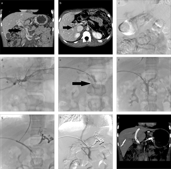
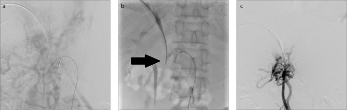
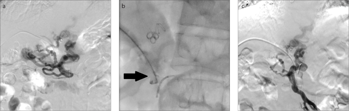
Purpose: We aimed to evaluate the feasibility and safety of a modified technique for portal vein recanalization, percutaneous transluminal sharp recanalization (PTSR), when performing transjugular intrahepatic portosystemic shunt (TIPS) for the treatment of chronic portal vein occlusion (CPVO) and portal hypertension.

Methods: Nine consecutive patients with CPVO and portal hypertension had undergone TIPS and PTSR procedure after failing in conventional percutaneous catheterization from March 2017 to July 2019. Technical success rates, effectiveness, and complications were evaluated. Follow-up of patients' clinical outcomes and shunt patency were performed periodically. Primary and secondary shunt patency were analyzed by Kaplan-Meier method.

Results: The occluded portal veins were successfully recanalized after failing in conventional percutaneous catheterization, and TIPS procedures were completed in all 9 patients. Two patients suffered from procedure-related complications. A portosystemic pressure gradient <12 mmHg, or a percent reduction of 25% to 50% of baseline, was achieved in all 9 patients after TIPS. During the median follow-up period of 28 months (range, 9-36 months), 1 patient experienced recurrent ascites and the other 8 patients remained asymptomatic. The cumulative rates of primary and secondary shunt patency were 66.67% and 100%, respectively, at 2 years.

Conclusion: As a supplementary method, PTSR is a feasible and safe method for portal vein recanalization when performing TIPS for patients with CPVO and portal hypertension.


Conflict of interest statement

Conflict of interest disclosure

The authors declared no conflicts of interest.

Figures

Figure 1. a–i
Figure 1. a–i
A 14-year-old boy (Patient 2) with recurrent variceal bleeding, who underwent splenectomy 8 years ago due to cryptogenic splenomegaly and hypersplenism. CT images (a, b) demonstrate complete main portal vein occlusion with portal-portal collateral veins (black arrow) and patent superior mesenteric vein. Indirect portography (c) shows complete occlusion of the main portal vein. Image (d) shows peripheral branch of right portal vein accessed transhepatically and conventional percutaneous catheterization failed in recanalization. Image (e) shows the patent superior mesenteric vein punctured through the residual trunk of the occluded portal vein by a 20-gauge Chiba needle (black arrow) under indirect portography and catheter positioning guidance. Image (f) shows successful access to superior mesenteric vein as identified by direct portography. Image (g) shows portal vein punctured via transjugular approach after variceal embolization, with the contrast-filled balloon as a target. Image (h) shows venogram obtained after shunt creation with a covered stent implantation demonstrating shunt patency. CT image (i) demonstrates shunt patency after the procedure.
Figure 2. a–c
Figure 2. a–c
A 44-year-old man (Patient 3) who suffered from ascites and protein S deficiency. Indirect portography (a) shows complete occlusion of the main portal vein. In image (b), under the indirect portography and catheter positioning guidance, the patent superior mesenteric vein was punctured through the residual trunk of the occluded portal vein by a 20-gauge Chiba needle (black arrow). Image (c) shows successful access to superior mesenteric vein as identified by direct portography.
Figure 3. a–c
Figure 3. a–c
A 52-year-old man (Patient 1) with recurrent variceal bleeding, who underwent liver transplantation and splenectomy 6 years ago due to cirrhosis. Direct portography via the transhepatic approach (a) shows complete occlusion of the main portal vein and conventional percutaneous catheterization failed in recanalization. In image (b), under the indirect portography and catheter positioning guidance, the patent superior mesenteric vein was punctured through the residual trunk of the occluded portal vein by a 20-gauge Chiba needle (black arrow). Image (c) shows successful access to superior mesenteric vein as identified by direct portography.

Similar articles

Cited by

References

    1. European Association for the Study of the Liver. EASL Clinical Practice Guidelines: Vascular diseases of the liver. J Hepatol. 2016;64:179–202. doi: 10.1016/j.jhep.2015.07.040. - DOI - PubMed
    1. Garcia-Pagán JC, Hernández-Guerra M, Bosch J. Extrahepatic portal vein thrombosis. Semin Liver Dis. 2008;28:282–292. doi: 10.1055/s-0028-1085096. - DOI - PubMed
    1. Wu M, Schuster M, Tadros M. Update on management of portal vein thrombosis and the role of novel anticoagulants. J Clin Transl Hepatol. 2019;7:154–164. doi: 10.14218/JCTH.2018.00057. - DOI - PMC - PubMed
    1. Senzolo M, Tibbals J, Cholongitas E, Triantos CK, Burroughs AK, Patch D. Transjugular intrahepatic portosystemic shunt for portal vein thrombosis with and without cavernous transformation. Aliment Pharmacol Ther. 2006;23:767–775. doi: 10.1111/j.1365-2036.2006.02820.x. - DOI - PubMed
    1. Van Ha TG, Hodge J, Funaki B, et al. Transjugular intrahepatic portosystemic shunt placement in patients with cirrhosis and concomitant portal vein thrombosis. Cardiovasc Intervent Radiol. 2006;29:785–790. doi: 10.1007/s00270-005-0090-4. - DOI - PubMed

LinkOut - more resources