. 2020 Sep 26:(163):10.3791/61657.
doi: 10.3791/61657.

Ultra-High-Speed Western Blot using Immunoreaction Enhancing Technology

Affiliations

Ultra-High-Speed Western Blot using Immunoreaction Enhancing Technology

Sayuri L Higashi et al. J Vis Exp. .

Abstract

A western blot (also known as an immunoblot) is a canonical method for biomedical research. It is commonly used to determine the relative size and abundance of specific proteins as well as post-translational protein modifications. This technique has a rich history and remains in widespread use due to its simplicity. However, the western blotting procedure famously takes hours, even days, to complete, with a critical bottleneck being the long incubation times that limit its throughput. These incubation steps are required due to the slow diffusion of antibodies from the bulk solution to the immobilized antigens on the membrane: the antibody concentration near the membrane is much lower than the bulk concentration. Here, we present an innovation that dramatically reduces these incubation intervals by improving antigen binding via cyclic draining and replenishing (CDR) of the antibody solution. We also utilized an immunoreaction enhancing technology to preserve the sensitivity of the assay. A combination of the CDR method with a commercial immunoreaction enhancing agent boosted the output signal and substantially reduced the antibody incubation time. The resulting ultra-high-speed western blot can be accomplished in 20 minutes without any loss in sensitivity. This method can be applied to western blots using both chemiluminescent and fluorescent detection. This simple protocol allows researchers to better explore the analysis of protein expression in many samples.


Conflict of interest statement

DISCLOSURES:

The authors have declared no competing interests directly relevant to the contents of this article.

Figures
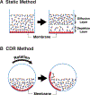
Figure 1
Figure 1
Schematic illustration of the cyclic draining-replenishing (CDR) method A. A depletion layer near the membrane rapidly forms under static conditions when a probe has a high affinity for the antigen but a limited ability to diffuse through the solution. Thus, travel of the probe to the membrane becomes a rate limiting step and long incubation times compensate for mass transfer limitation. B. CDR method by rotating the tube while the membrane adheres to the wall eliminates the depletion layer and replenishes the same probe solution to keep the probe concentration near the membrane constant. This figure has been modified from Higashi et al.
Figure 2
Figure 2
A. Different amounts of 293 cell lysates (1:2 serial dilutions from 8.8 μg/lane) were separated by SDS-PAGE, followed by transfer to a PVDF membrane and blocking with 5 % skim milk in PBS-T. The blot was probed with mouse anti-β-actin antibody (1:3,000 dilution). B. After separation of conditioned media derived from transfected 293 cells with pAPTAG5 (GenHunter) containing the secreted (His)6 tagged alkaline phosphatase (AP, 8 × 10⁻¹⁴ mol/lane), proteins were transferred to PVDF membranes. Each membrane was subjected to western blot with different concentrations of anti-6X His tag antibody (1:2 serial dilutions from 400 ng/ml). The membranes were imaged as a single image and dotted lines indicate the border of individual membranes. This figure has been modified from Higashi et al.
Figure 3
Figure 3
Simultaneous detection of multiple targets on a western blot using fluorescent detection and CDR in conjunction with IRE Different amounts of lysates (1:2 serial dilutions from 10 μg/lane) from 293 cells transfected with pAPTAG5 were separated and transferred to a PVDF membrane. After blocking with the Blocking Buffer for fluorescent detection (BFD) for 1 h, the blot was probed with anti-6X His tag and anti β-actin antibodies, followed by IRDye 800CW goat anti-rabbit IgG and IRDye 680RD goat anti-mouse IgG. A: antibodies diluted with 10 % IRE solution under CDR condition, B: antibodies diluted with BFD containing 0.1 % Tween20 under static condition. Because of higher sensitivity with CDR and IRE combination, the degradation of (His)6 tagged AP was seen. This figure has been modified from Higashi et al.
Figure 4
Figure 4
Schematic illustration of an enhanced and ultra-high speed CDR western blot compared to a traditional static method. This figure has been modified from Higashi et al.

References

    1. MacPhee DJ Methodological considerations for improving Western blot analysis. Journal of Pharmacol Toxicol Methods. 61 (2), 171–177, doi:10.1016/j.vascn.2009.12.001, (2010). - DOI - PubMed
    1. Janes KA An analysis of critical factors for quantitative immunoblotting. Sci Signal. 8 (371), rs2, doi:10.1126/scisignal.2005966, (2015). - DOI - PMC - PubMed
    1. Schuck P & Zhao H The role of mass transport limitation and surface heterogeneity in the biophysical characterization of macromolecular binding processes by SPR biosensing. Methods in Molecular Biology. 627 15–54, doi:10.1007/978-1-60761-670-2_2, (2010). - DOI - PMC - PubMed
    1. Li J, Zrazhevskiy P & Gao X Eliminating Size-Associated Diffusion Constraints for Rapid On-Surface Bioassays with Nanoparticle Probes. Small. 12 (8), 1035–1043, doi:10.1002/smll.201503101, (2016). - DOI - PMC - PubMed
    1. TOYOBO. IMMUNOREACTION ENHANCING: IE TECHNOLOGY. http://www.toyobousa.com/lifescience-immunoreaction-enhancing.html

Publication types

LinkOut - more resources