Review
. 2021 Aug;36(8):2189-2201.
doi: 10.1007/s00467-020-04756-4. Epub 2020 Oct 12.

Childhood HIV-associated nephropathy: 36 years later

Affiliations
Review

Childhood HIV-associated nephropathy: 36 years later

Patricio E Ray et al. Pediatr Nephrol. 2021 Aug.

Abstract

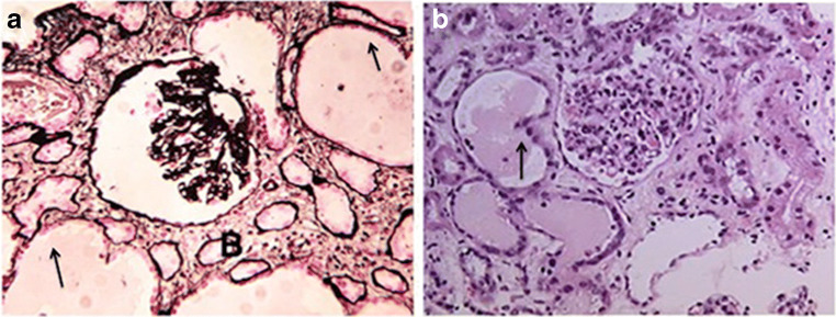
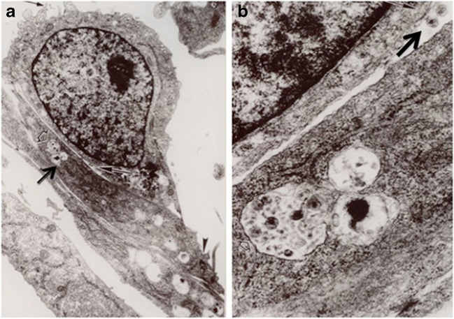
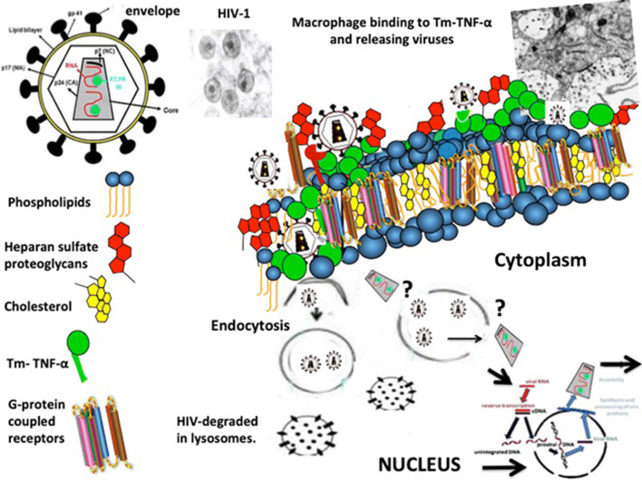
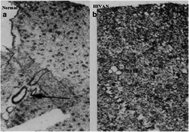
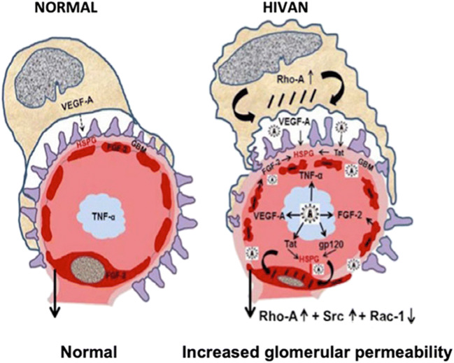
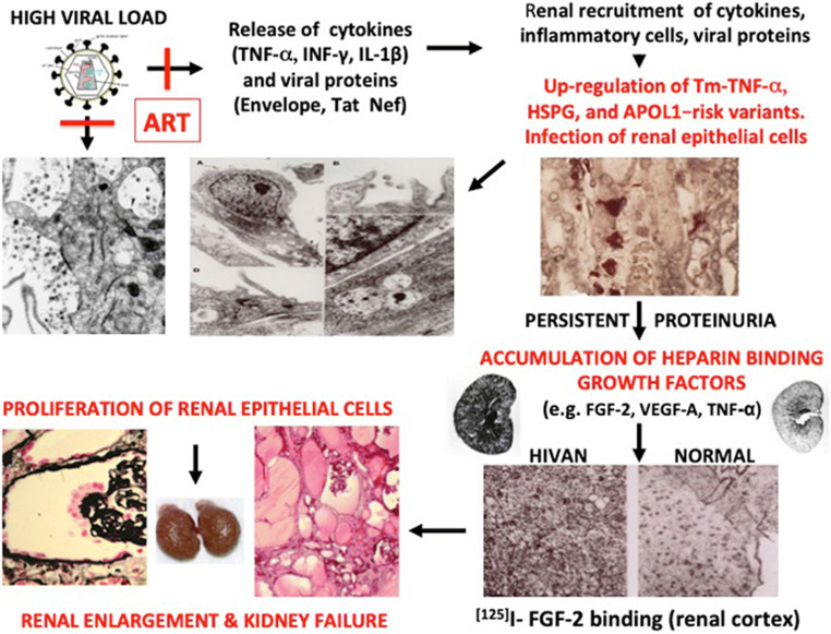
HIV-associated nephropathy (HIVAN) predominantly affects people of African ancestry living with HIV who do not receive appropriate antiretroviral therapy (ART). Childhood HIVAN is characterized by heavy proteinuria and decreased kidney function. Kidney histology shows mesangial expansion, classic or collapsing glomerulosclerosis, and microcystic renal tubular dilatation leading to kidney enlargement. The pathogenesis of HIVAN involves the kidney recruitment of inflammatory cells and the infection of kidney epithelial cells. In addition, both viral and genetic factors play key roles in this disease. Modern ART has improved the outcome and decreased the prevalence of childhood HIVAN. However, physicians have had modest success providing chronic ART to children and adolescents, and we continue to see children with HIVAN all over the world. This article discusses the progress made during the last decade in our understanding of the pathogenesis and treatment of childhood HIVAN, placing particular emphasis on the mechanisms that mediate the infection of kidney epithelial cells, and the roles of cytokines, the HIV-Tat gene, and the Apolipoprotein-1 (APOL1) gene risk variants in this disease. In view of the large number of children living with HIV at risk of developing HIVAN, better prevention and treatment programs are needed to eradicate this disease.

Keywords: APOL-1; Antiretroviral therapy; HIV nephropathy; HIV-Tat; Heparan sulfate proteoglycans; Infection of podocytes; Kidney epithelial cells; Pediatric AIDS.


Conflict of interest statement

The authors declare that they have no conflict of interest.

Figures

Fig. 1
Fig. 1
Panels (a) and (b) show representative kidney sections from children with HIVAN. Panel (a) shows a kidney biopsy with collapsing glomerulopathy stained with period acid Schiff. Panel (b) shows a kidney biopsy with mesangial expansion and enlarged glomeruli stained with H&E. Both panels show the tubular microcystic changes characteristic of childhood HIVAN (arrows). Original magnification × 200
Fig. 2
Fig. 2
Panels (a) and (b) show a mononuclear cell on top of renal tubular epithelial cells (RTEC) cultured from a child with HIVAN. The arrowhead in panel (a) points to a tight junction between RTEc. The arrow points to HIV viral particles inside RTEc. Panel (b) shows a higher magnification of the viral particles inside RTEc, as well as viral particles in the space between mononuclear and RTEc (arrow). Original magnification ×10,000 (a) and ×50,000 (b)
Fig. 3
Fig. 3
Proposed mechanisms for explaining the infection of kidney epithelial cells cultured from children with HIVAN. Transmembrane-TNF-α (Tm-TNF-α) expressed on the surface of kidney epithelial and mononuclear cells facilitates the binding of HIV-infected mononuclear cells to kidney epithelial cells. Viruses are released into a “virological synapse” created between these cells (Fig. 2). HIV is internalized via an envelope-clathrin-mediated endocytosis mechanism prior to membrane fusion. Heparan sulfate proteoglycans facilitate the binding and entry of HIV-1 via endocytosis. Once inside endosome-like compartments, viral particles can be degraded in endolysosomal compartments (non-productive infection). A small number of virus particles may undergo fusion in endosome-like compartments or escape into the cytosol through other mechanisms that are not clearly understood at the present time (?). Viral particles released into the cytoplasm undergo reverse transcription and integration into the nucleus. In turn, after undergoing several assembling steps, new infectious viral particles can be released
Fig. 4
Fig. 4
Total binding of 125I-Fibroblast Growth Factor-2 to heparan sulfated proteoglycans (HSPG) located in glomeruli, vessels, and the kidney interstitium of children with and without HIVAN. Panel (a) shows a representative kidney section from a control child (normal). The arrow in panel (a) points to 125I-FGF binding to kidney vessels. Panel (b) shows increased binding of 125I-FGF to HSPG in the kidney cortical interstitium in a child with HIVAN. Original magnification ×60. (Figure is reproduced with permission of Springer from Ray PE, Liu Xu, Rakusan T, Liu Xue-Hui. “A 20-year history of childhood HIV-associated nephropathy” Pediatric Nephrology 19: 1075–1092, page 1083; License Number 3984431229899)
Fig. 5
Fig. 5
HIV-Tat, fibroblast growth factor-2 (FGF-2), and vascular endothelial growth factor (VEGF-A) increase the permeability of the glomerular filtration barrier. Under normal conditions, VEGF-A released from podocytes is needed to maintain the normal structure and survival of endothelial cells. FGF-2 is not released from healthy endothelial cells, and very low levels of TNF-α are released from mononuclear cells. In contrast, during the early stages of HIVAN, HIV-Tat and other heparin binding cytokines or viral proteins released by HIV-infected mononuclear cells, injured endothelial cells, or podocytes affect the phosphorylation and activity of Rho-A, Src, and Rac-1 in podocytes and glomerular endothelial cells. These changes alter the cytoskeleton structure of podocytes and glomerular endothelial cells and increase the permeability of the glomerular filtration barrier causing proteinuria
Fig. 6
Fig. 6
Proposed mechanisms for explaining the pathogenesis of childhood HIVAN. A high viral load increases the release of cytokines and viral proteins in the circulation. Cytokines, viral proteins, and inflammatory cells are recruited in the kidney, increasing the expression of Tm-TNF-α, heparan sulfate proteoglycans, and APOL1 in kidney epithelial, endothelial, and tubulointerstitial cells. All these changes induce glomerular and tubulointerstitial injury, increasing the recruitment of inflammatory cells, and facilitating the infection and injury of more kidney epithelial cells causing persistent proteinuria. The accumulation of heparin binding growth factors and viral proteins increase the proliferation of podocytes and tubular epithelial cells leading to kidney enlargement and kidney failure. Antiretroviral therapy (ATR) blocks the early stages and development of HIVAN, but may not affect the secondary proliferative changes once the kidney damage is established

References

    1. Rao TK, Filippone EJ, Nicastri AD, Landesman SH, Frank E, Chen CK, Friedman EA. Associated focal and segmental glomerulosclerosis in the acquired immunodeficiency syndrome. N Engl J Med. 1984;310:669–673. - PubMed
    1. Strauss J, Abitbol C, Zilleruelo G, Scott G, Paredes A, Malaga S, Montane B, Mitchell C, Parks W, Pardo V. Renal disease in children with the acquired immunodeficiency syndrome. N Engl J Med. 1989;321:625–630. - PubMed
    1. Ray PE, Rakusan T, Loechelt BJ, Selby DM, Liu XH, Chandra RS. Human immunodeficiency virus (HIV)-associated nephropathy in children from the Washington, D.C. area: 12 years’ experience. Semin Nephrol. 1998;18:396–405. - PubMed
    1. McCulloch MI, Ray PE. Kidney disease in HIV-positive children. Semin Nephrol. 2008;28:585–594. - PMC - PubMed
    1. Children, HIV and AIDS. Global Snapshot 2019. https://data.unicef.org/resources/children-hiv-aids-global-snapshot/data.unicef.org