

Steps to use artificial intelligence in echocardiography
- PMID: 33044715
- PMCID: PMC7549428
- DOI: 10.1007/s12574-020-00496-4
Steps to use artificial intelligence in echocardiography
Abstract
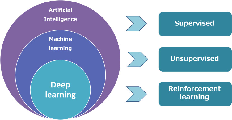
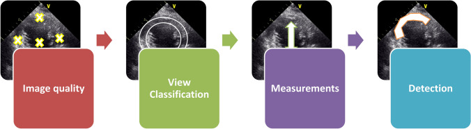
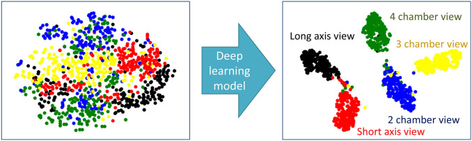
Artificial intelligence (AI) has influenced every field of cardiovascular imaging in all phases from acquisition to reporting. Compared with computed tomography and magnetic resonance imaging, there is an issue of high observer variation in the interpretation of echocardiograms. Therefore, AI can help minimize the observer variation and provide accurate diagnosis in the field of echocardiography. In this review, we summarize the necessity for automated diagnosis in the echocardiographic field, and discuss the results of AI application to echocardiography and future perspectives. Currently, there are two roles for AI in cardiovascular imaging. One is the automation of tasks performed by humans, such as image segmentation, measurement of cardiac structural and functional parameters. The other is the discovery of clinically important insights. Most reported applications were focused on the automation of tasks. Moreover, algorithms that can obtain cardiac measurements are also being reported. In the next stage, AI can be expected to expand and enrich existing knowledge. With the continual evolution of technology, cardiologists should become well versed in this new knowledge of AI and be able to harness it as a tool. AI can be incorporated into everyday clinical practice and become a valuable aid for many healthcare professionals dealing with cardiovascular diseases.
Keywords: Artificial intelligence; Cardiovascular imaging; Deep learning; Echocardiography; Radiomic.
Conflict of interest statement
Kenya Kusunose declares that they have no conflict of interest.
Figures


References
-
- LeCun Y, Bengio Y, Hinton G. Deep Learning Nat. 2015;521:436–444. - PubMed
Publication types
MeSH terms
LinkOut - more resources
Full Text Sources
