Meta-Analysis
. 2022 Jan;43(1):470-499.
doi: 10.1002/hbm.25204. Epub 2020 Oct 12.

Greater male than female variability in regional brain structure across the lifespan

Lara M Wierenga  1   2 Gaelle E Doucet  3   4 Danai Dima  5   6 Ingrid Agartz  7   8   9 Moji Aghajani  10   11   12 Theophilus N Akudjedu  13   14 Anton Albajes-Eizagirre  15   16   17 Dag Alnaes  7   18 Kathryn I Alpert  19 Ole A Andreassen  7   18 Alan Anticevic  20 Philip Asherson  21 Tobias Banaschewski  22 Nuria Bargallo  23   24 Sarah Baumeister  22 Ramona Baur-Streubel  25 Alessandro Bertolino  26 Aurora Bonvino  26 Dorret I Boomsma  27 Stefan Borgwardt  28   29 Josiane Bourque  30   31 Anouk den Braber  27   32 Daniel Brandeis  22   33   34   35 Alan Breier  36 Henry Brodaty  37   38 Rachel M Brouwer  39 Jan K Buitelaar  40   41 Geraldo F Busatto  42 Vince D Calhoun  43 Erick J Canales-Rodríguez  15   16 Dara M Cannon  13 Xavier Caseras  44 Francisco X Castellanos  45   46 Tiffany M Chaim-Avancini  42 Christopher Rk Ching  47 Vincent P Clark  48   49 Patricia J Conrod  31   50 Annette Conzelmann  51   52 Fabrice Crivello  53 Christopher G Davey  54   55 Erin W Dickie  56   57 Stefan Ehrlich  58 Dennis Van't Ent  27 Simon E Fisher  59   60 Jean-Paul Fouche  61 Barbara Franke  60   62   63 Paola Fuentes-Claramonte  15   16 Eco Jc de Geus  27 Annabella Di Giorgio  64 David C Glahn  65   66 Ian H Gotlib  67 Hans J Grabe  68   69 Oliver Gruber  70 Patricia Gruner  20 Raquel E Gur  30   71 Ruben C Gur  30 Tiril P Gurholt  7   18 Lieuwe de Haan  72 Beathe Haatveit  7   18 Ben J Harrison  73 Catharina A Hartman  74 Sean N Hatton  75   76 Dirk J Heslenfeld  77 Odile A van den Heuvel  10   78 Ian B Hickie  75 Pieter J Hoekstra  79 Sarah Hohmann  22 Avram J Holmes  20   80   81 Martine Hoogman  60   62 Norbert Hosten  82 Fleur M Howells  83   84 Hilleke E Hulshoff Pol  39 Chaim Huyser  85   86 Neda Jahanshad  47 Anthony C James  87   88 Jiyang Jiang  37 Erik G Jönsson  7   9 John A Joska  84 Andrew J Kalnin  89 Karolinska Schizophrenia Project (KaSP) ConsortiumMarieke Klein  39   60   62 Laura Koenders  72 Knut K Kolskår  18   90   91 Bernd Krämer  70 Jonna Kuntsi  21 Jim Lagopoulos  92   93 Luisa Lazaro  16   94   95   96 Irina S Lebedeva  97 Phil H Lee  81   98 Christine Lochner  99 Marise Wj Machielsen  100 Sophie Maingault  101 Nicholas G Martin  102 Ignacio Martínez-Zalacaín  103   104 David Mataix-Cols  9 Bernard Mazoyer  105   106 Brenna C McDonald  107 Colm McDonald  13 Andrew M McIntosh  108 Katie L McMahon  109   110 Genevieve McPhilemy  13 Dennis van der Meer  7   18   111 José M Menchón  16   103   104 Jilly Naaijen  40 Lars Nyberg  112   113 Jaap Oosterlaan  114   115 Yannis Paloyelis  6 Paul Pauli  116   117 Giulio Pergola  26   118 Edith Pomarol-Clotet  15   16 Maria J Portella  16   119 Joaquim Radua  9   15   16   17   120 Andreas Reif  121 Geneviève Richard  7   18 Joshua L Roffman  122 Pedro Gp Rosa  42 Matthew D Sacchet  123 Perminder S Sachdev  37   124 Raymond Salvador  15   16 Salvador Sarró  15   16 Theodore D Satterthwaite  30 Andrew J Saykin  107   125 Mauricio H Serpa  42 Kang Sim  126   127 Andrew Simmons  128 Jordan W Smoller  81   129 Iris E Sommer  130 Carles Soriano-Mas  16   103   131 Dan J Stein  132 Lachlan T Strike  133 Philip R Szeszko  3   134 Henk S Temmingh  84 Sophia I Thomopoulos  47 Alexander S Tomyshev  97 Julian N Trollor  37 Anne Uhlmann  84   135 Ilya M Veer  136 Dick J Veltman  137 Aristotle Voineskos  56 Henry Völzke  138   139   140 Henrik Walter  136 Lei Wang  19 Yang Wang  141 Bernd Weber  142 Wei Wen  37 John D West  107 Lars T Westlye  7   18   90 Heather C Whalley  108   143 Steven Cr Williams  144 Katharina Wittfeld  68   69 Daniel H Wolf  30 Margaret J Wright  133   145 Yuliya N Yoncheva  146 Marcus V Zanetti  42   147 Georg C Ziegler  148 Greig I de Zubicaray  110 Paul M Thompson  47 Eveline A Crone  1   2   149 Sophia Frangou  3   150 Christian K Tamnes  7   8   151
Affiliations
Meta-Analysis

Greater male than female variability in regional brain structure across the lifespan

Lara M Wierenga et al. Hum Brain Mapp. 2022 Jan.

Abstract

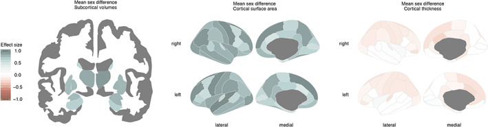
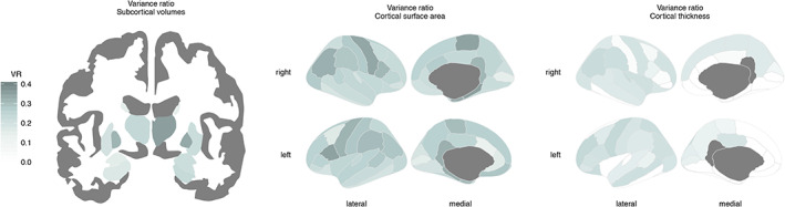
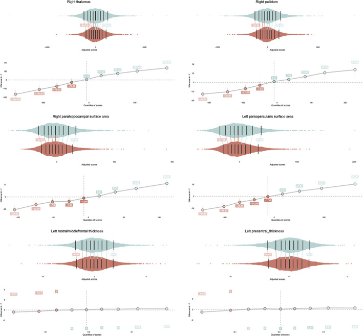
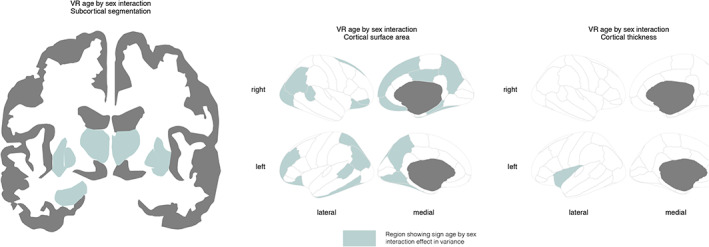
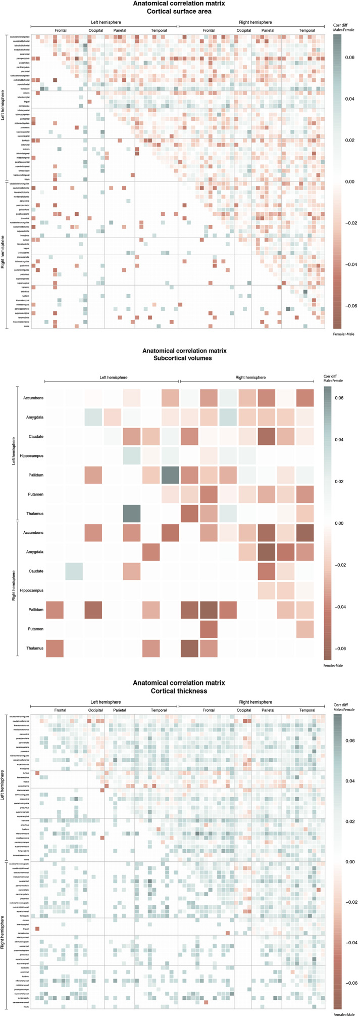
For many traits, males show greater variability than females, with possible implications for understanding sex differences in health and disease. Here, the ENIGMA (Enhancing Neuro Imaging Genetics through Meta-Analysis) Consortium presents the largest-ever mega-analysis of sex differences in variability of brain structure, based on international data spanning nine decades of life. Subcortical volumes, cortical surface area and cortical thickness were assessed in MRI data of 16,683 healthy individuals 1-90 years old (47% females). We observed significant patterns of greater male than female between-subject variance for all subcortical volumetric measures, all cortical surface area measures, and 60% of cortical thickness measures. This pattern was stable across the lifespan for 50% of the subcortical structures, 70% of the regional area measures, and nearly all regions for thickness. Our findings that these sex differences are present in childhood implicate early life genetic or gene-environment interaction mechanisms. The findings highlight the importance of individual differences within the sexes, that may underpin sex-specific vulnerability to disorders.


Conflict of interest statement

The authors declare the following competing interests: OAA: Speaker's honorarium from Lundbeck, Consultant of HealthLyti; PA: Received payments for consultancy to Shire/Takeda, Medic, educational/research awards from Shire/Takeda, GW Pharma, Janssen‐Cila, speaker at sponsored events for Shire, Flynn Pharma, Medic; TB: advisory or consultancy role for Lundbeck, Medice, Neurim Pharmaceuticals, Oberberg GmbH, Shire, and Infectopharm, conference support or speaker's fee by Lilly, Medice, and Shire, received royalities from Hogrefe, Kohlhammer, CIP Medien, Oxford University Press ‐ the present work is unrelated to the above grants and relationship; DB: serves as an unpaid scientific consultant for an EU‐funded neurofeedback trial that is unrelated to the present work; HB: Advisory Board, Nutricia Australi; CRKC: received partial research support from Biogen, Inc. (Boston, USA) for work unrelated to the topic of this manuscript; BF: received educational speaking fees from Medice; HJG: received travel grants and speakers honoraria from Fresenius Medical Care, Neuraxpharm, Servier and Janssen Cilag as well as research funding from Fresenius Medical Care; NJ and PMT: MPI of a research related grant from Biogen, Inc., for research unrelated to the contents of this manuscript; JK: given talks at educational events sponsored by Medic; all funds are received by King's College London and used for studies of ADHD; DM‐C: receives fees from UpToDate, Inc and Elsevier, all unrelated to the current work; AMM: received research support from Eli Lilly, Janssen, and the Sackler Foundation, and speaker fees from Illumina and Janssen; DJS: received research grants and/or honoraria from Lundbeck and Sun. The remaining authors declare no competing interests.

Figures

FIGURE 1
FIGURE 1
Sex differences in volumetric measures of subcortical volumes (left), cortical surface area (center), and cortical thickness (right). Shown are effect sizes (Cohen's d‐value) of FDR corrected mean sex differences. Greater mean values for males are displayed in blue, greater mean values for females are displayed in red. Darker colors indicate larger effect sizes
FIGURE 2
FIGURE 2
Sex differences in variance ratio for subcortical volumes (Left), cortical surface area (center), and cortical thickness (right). Shown are log transformed variance ratios, where significant larger variance ratio for males than females is displayed in blue ranging from 0 to 1. Darker colors indicate a larger variance ratio
FIGURE 3
FIGURE 3
Jittered marginal distribution scatterplots are displayed together with their shift function for the top three variance ratio effects of subcortical volumes (top), cortical surface area (middle) and cortical thickness (right). The central, darkest line on each distribution is the median, note that main sex effects are removed. The other lines mark the deciles of each distribution. The shift values are included, which refer to the number of units that the male (upper) distribution would have to be shifted to match the female (lower) distribution. Confidence intervals are included for each of these shift values
FIGURE 4
FIGURE 4
Regions where sex differences in variability of brain structure interacted with age displayed for subcortical volumes (left), cortical surface area (center), and cortical thickness (right)
FIGURE 5
FIGURE 5
Sex differences in variability interacted with age in 50% of the subcortical volumes, 30% of the surface area measures, and only one thickness measure. Three representative results are shown: right thalamus volume (top left), surface area of the right parahippocampal gyrus (top right) and thickness of the left insula (bottom center). Absolute residual values are modeled across the age range. Effects showed larger male than female variance in the younger age group, this effect attenuated with increasing age
FIGURE 6
FIGURE 6
(a–c) Stronger anatomical correlations for males than females are indicated in blue (larger homogeneity in males than females), while stronger correlations for females are displayed in red (larger homogeneity in females than males). The bottom left half shows the significant variance ratio's only, using two sided permutation testing. Results are displayed for subcortical volumes (a), surface area (b), and cortical thickness (c). Cortical regions are ordered by lobe and hemisphere (left frontal, left occipital, left parietal, left temporal, right frontal, right occipital, right parietal, right temporal)

Similar articles

  • Cortical and Subcortical Brain Morphometry Differences Between Patients With Autism Spectrum Disorder and Healthy Individuals Across the Lifespan: Results From the ENIGMA ASD Working Group.
    van Rooij D, Anagnostou E, Arango C, Auzias G, Behrmann M, Busatto GF, Calderoni S, Daly E, Deruelle C, Di Martino A, Dinstein I, Duran FLS, Durston S, Ecker C, Fair D, Fedor J, Fitzgerald J, Freitag CM, Gallagher L, Gori I, Haar S, Hoekstra L, Jahanshad N, Jalbrzikowski M, Janssen J, Lerch J, Luna B, Martinho MM, McGrath J, Muratori F, Murphy CM, Murphy DGM, O'Hearn K, Oranje B, Parellada M, Retico A, Rosa P, Rubia K, Shook D, Taylor M, Thompson PM, Tosetti M, Wallace GL, Zhou F, Buitelaar JK. van Rooij D, et al. Am J Psychiatry. 2018 Apr 1;175(4):359-369. doi: 10.1176/appi.ajp.2017.17010100. Epub 2017 Nov 17. Am J Psychiatry. 2018. PMID: 29145754 Free PMC article.
  • Sex Differences in Variability of Brain Structure Across the Lifespan.
    Forde NJ, Jeyachandra J, Joseph M, Jacobs GR, Dickie E, Satterthwaite TD, Shinohara RT, Ameis SH, Voineskos AN. Forde NJ, et al. Cereb Cortex. 2020 Sep 3;30(10):5420-5430. doi: 10.1093/cercor/bhaa123. Cereb Cortex. 2020. PMID: 32483605 Free PMC article.
  • Association of Copy Number Variation of the 15q11.2 BP1-BP2 Region With Cortical and Subcortical Morphology and Cognition.
    Writing Committee for the ENIGMA-CNV Working Group; van der Meer D, Sønderby IE, Kaufmann T, Walters GB, Abdellaoui A, Ames D, Amunts K, Andersson M, Armstrong NJ, Bernard M, Blackburn NB, Blangero J, Boomsma DI, Brodaty H, Brouwer RM, Bülow R, Cahn W, Calhoun VD, Caspers S, Cavalleri GL, Ching CRK, Cichon S, Ciufolini S, Corvin A, Crespo-Facorro B, Curran JE, Dalvie S, Dazzan P, de Geus EJC, de Zubicaray GI, de Zwarte SMC, Delanty N, den Braber A, Desrivieres S, Di Forti M, Doherty JL, Donohoe G, Ehrlich S, Eising E, Espeseth T, Fisher SE, Fladby T, Frei O, Frouin V, Fukunaga M, Gareau T, Glahn DC, Grabe HJ, Groenewold NA, Gústafsson Ó, Haavik J, Haberg AK, Hashimoto R, Hehir-Kwa JY, Hibar DP, Hillegers MHJ, Hoffmann P, Holleran L, Hottenga JJ, Hulshoff Pol HE, Ikeda M, Jacquemont S, Jahanshad N, Jockwitz C, Johansson S, Jönsson EG, Kikuchi M, Knowles EEM, Kwok JB, Le Hellard S, Linden DEJ, Liu J, Lundervold A, Lundervold AJ, Martin NG, Mather KA, Mathias SR, McMahon KL, McRae AF, Medland SE, Moberget T, Moreau C, Morris DW, Mühleisen TW, Murray RM, Nordvik JE, Nyberg L, Olde Loohuis LM, Ophoff RA, Owen MJ, Paus T, Pausova Z, Peralta JM, Pike B, Prieto C, Quinlan EB, Reinbold CS,… See abstract for full author list ➔ Writing Committee for the ENIGMA-CNV Working Group, et al. JAMA Psychiatry. 2020 Apr 1;77(4):420-430. doi: 10.1001/jamapsychiatry.2019.3779. JAMA Psychiatry. 2020. PMID: 31665216 Free PMC article.
  • What we learn about bipolar disorder from large-scale neuroimaging: Findings and future directions from the ENIGMA Bipolar Disorder Working Group.
    Ching CRK, Hibar DP, Gurholt TP, Nunes A, Thomopoulos SI, Abé C, Agartz I, Brouwer RM, Cannon DM, de Zwarte SMC, Eyler LT, Favre P, Hajek T, Haukvik UK, Houenou J, Landén M, Lett TA, McDonald C, Nabulsi L, Patel Y, Pauling ME, Paus T, Radua J, Soeiro-de-Souza MG, Tronchin G, van Haren NEM, Vieta E, Walter H, Zeng LL, Alda M, Almeida J, Alnaes D, Alonso-Lana S, Altimus C, Bauer M, Baune BT, Bearden CE, Bellani M, Benedetti F, Berk M, Bilderbeck AC, Blumberg HP, Bøen E, Bollettini I, Del Mar Bonnin C, Brambilla P, Canales-Rodríguez EJ, Caseras X, Dandash O, Dannlowski U, Delvecchio G, Díaz-Zuluaga AM, Dima D, Duchesnay É, Elvsåshagen T, Fears SC, Frangou S, Fullerton JM, Glahn DC, Goikolea JM, Green MJ, Grotegerd D, Gruber O, Haarman BCM, Henry C, Howells FM, Ives-Deliperi V, Jansen A, Kircher TTJ, Knöchel C, Kramer B, Lafer B, López-Jaramillo C, Machado-Vieira R, MacIntosh BJ, Melloni EMT, Mitchell PB, Nenadic I, Nery F, Nugent AC, Oertel V, Ophoff RA, Ota M, Overs BJ, Pham DL, Phillips ML, Pineda-Zapata JA, Poletti S, Polosan M, Pomarol-Clotet E, Pouchon A, Quidé Y, Rive MM, Roberts G, Ruhe HG, Salvador R, Sarró S, Satterthwaite TD, Schene AH, Sim K, Soares JC, Stäblein M, Stein D… See abstract for full author list ➔ Ching CRK, et al. Hum Brain Mapp. 2022 Jan;43(1):56-82. doi: 10.1002/hbm.25098. Epub 2020 Jul 29. Hum Brain Mapp. 2022. PMID: 32725849 Free PMC article. Review.
  • Mapping brain asymmetry in health and disease through the ENIGMA consortium.
    Kong XZ, Postema MC, Guadalupe T, de Kovel C, Boedhoe PSW, Hoogman M, Mathias SR, van Rooij D, Schijven D, Glahn DC, Medland SE, Jahanshad N, Thomopoulos SI, Turner JA, Buitelaar J, van Erp TGM, Franke B, Fisher SE, van den Heuvel OA, Schmaal L, Thompson PM, Francks C. Kong XZ, et al. Hum Brain Mapp. 2022 Jan;43(1):167-181. doi: 10.1002/hbm.25033. Epub 2020 May 18. Hum Brain Mapp. 2022. PMID: 32420672 Free PMC article. Review.

Cited by

References

    1. Alnæs, D. , Kaufmann, T. , van der Meer, D. , Córdova‐Palomera, A. , Rokicki, J. , Moberget, T. , … Westlye, L. T. (2019). Brain heterogeneity in schizophrenia and its association with polygenic risk. JAMA Psychiatry, 76(7), 739. - PMC - PubMed
    1. Arden, R. , & Plomin, R. (2006). Sex differences in variance of intelligence across childhood. Personality and Individual Differences, 41, 39–48.
    1. Arnold, A. P. (2012). The end of gonad‐centric sex determination in mammals. Trends in Genetics, 28(2), 55–61. - PMC - PubMed
    1. Aron, A. R. , Robbins, T. W. , & Poldrack, R. A. (2004). Inhibition and the right inferior frontal cortex. Trends in Cognitive Sciences, 8(4), 170–177. - PubMed
    1. Baaré, W. , Pol, H. , Boomsma, D. I. , & Posthuma, D. (2001). Quantitative genetic modeling of variation in human brain morphology. Cerebral Cortex, 111(9), 816–824. - PubMed

Publication types

Grants and funding