Review
. 2022 Jul:203:594-603.
doi: 10.1016/j.ymeth.2020.10.003. Epub 2020 Oct 10.

CRISPR-based assays for rapid detection of SARS-CoV-2

Affiliations
Review

CRISPR-based assays for rapid detection of SARS-CoV-2

Vivek S Javalkote et al. Methods. 2022 Jul.

Abstract

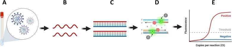
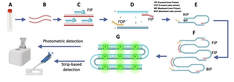
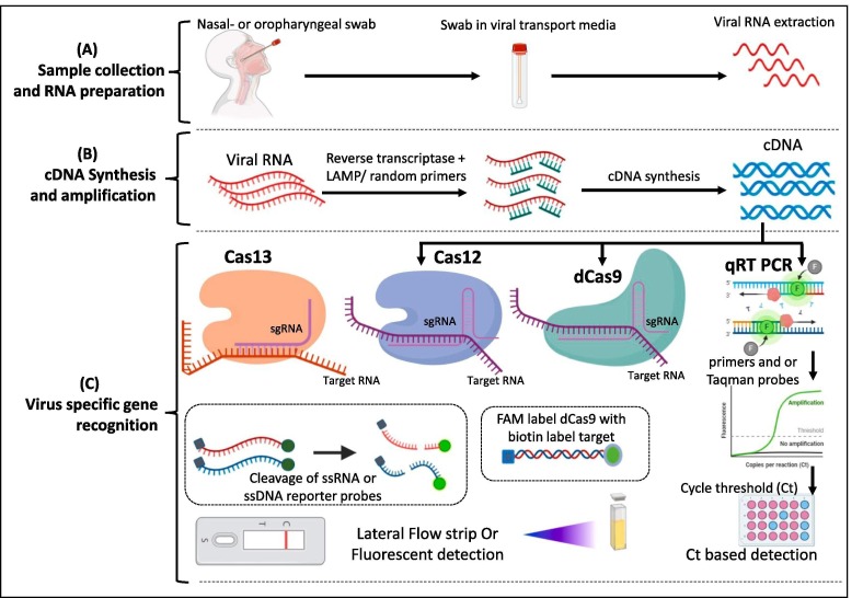
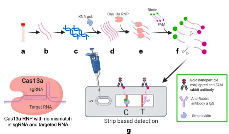
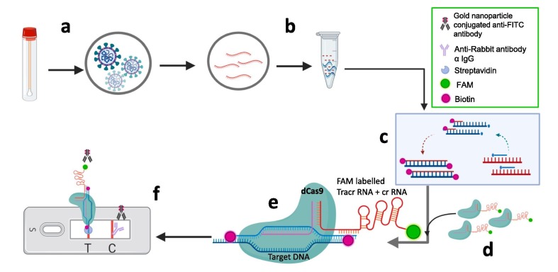
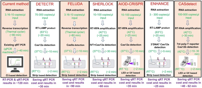
COVID-19 pandemic posed an unprecedented threat to global public health and economies. There is no effective treatment of the disease, hence, scaling up testing for rapid diagnosis of SARS-CoV-2 infected patients and quarantine them from healthy individuals is one the best strategies to curb the pandemic. Establishing globally accepted easy-to-access diagnostic tests is extremely important to understanding the epidemiology of the present pandemic. While nucleic acid based tests are considered to be more sensitive with respect to serological tests but present gold standard qRT-PCR-based assays possess limitations such as low sample throughput, requirement for sophisticated reagents and instrumentation. To overcome these shortcomings, recent efforts of incorporating LAMP-based isothermal detection, and minimizing the number of reagents required are on rise. CRISPR based novel techniques, when merge with isothermal and allied technologies, promises to provide sensitive and rapid detection of SARS-CoV-2 nucleic acids. Here, we discuss and present compilation of state-of-the-art detection techniques for COVID-19 using CRISPR technology which has tremendous potential to transform diagnostics and epidemiology.

Keywords: COVID-19; CRISPR diagnostics; Diagnostic advancements; Molecular testing; SARS-CoV-2.


Conflict of interest statement

The authors declare that they have no known competing financial interests or personal relationships that could have appeared to influence the work reported in this paper.

Figures

Fig. 1
Fig. 1
Flow diagram demonstrating steps of qRT PCR based detection of viral nucleic acid in sample. A) Nasopharyngeal swab collection from infected patients B) RNAs extracted from samples (red strand) C) Reverse transcription for cDNA synthesis (green strand) and amplification (RT-PCR) conducted by using target specific primers (eg. SARS-CoV-2N gene or RDRP gene) D) Quantitative real time PCR (qRT-PCR) conducted to identify positive samples.E) Final result. Amplification of sample monitored in real time based on fluorescence generated by incorporation of dyes like SYBR green in double stranded DNA. Further TaqMan probes specific to target sequence increases the specificity and sensitivity. (For interpretation of the references to color in this figure legend, the reader is referred to the web version of this article.)
Fig. 2
Fig. 2
Schematic representations of LAMP for detection of viruses in samples. A) Sample (Nasopharyngeal swab) collection, B) RNA extracted from samples (green strand), C) Reverse transcription for cDNA synthesis (red strand) and amplification by strand displacement reaction proceeds at a constant temperature, D - F) Shows LAMP reaction: Target sequence is amplified using four sets of primers with a highly efficient polymerase. Four primer used are specific to target regions involves Forward Inner Primer (FIP), Forward Outer Primer (FOP), Backward Inner Primer (BIP) and Backward Outer Primer (BOP). FOP amplifies the one strand of DNA and displaces the other strand. Displaced strand forms loop and serves as a template for BIP and complementary strand is formed. BOP amplifies the complementary strand and form a dumbbell shape structure. Amplification is achieved by repeating the extensions by respective primers. G) Amplified nucleic acids further can be detected by binding with fluorescent dye, qRT-PCR or paper strip based detection methods (Amplification also observed in real time by using pH sensitive dyes in reaction mixture and monitoring the color change. Due to accumulation of H + ions in the reaction mixture, which leads to increased pH). (For interpretation of the references to color in this figure legend, the reader is referred to the web version of this article.)
Fig. 3
Fig. 3
Fundamental steps of various CRISPR based COVID-19 detection tests. (A) Collection of sample, transport in Viral transport medium and isolation of RNA (B) Conversion of RNA to cDNA by Reverse transcription. This amplification step could be conducted at isothermal conditions by following RT-RPA of RT-LAMP method. (C) Amplified nucleic acid of viral sample need to be identified by specific viral gene amplification. Conventional method (qRT PCR) utilizes viral gene specific primers to amplify the specific gene segment and further these segments detected by double strand intercalating SYBR dye or fluorescent probes. Recent advancements in CRISPR based diagnostics (CRISPR-Dx) enabled utilization of different Cas enzymes for detection with high sensitivity either from RNA (Cas-13 based detection) or from cDNA (Cas12, Cas9 based etc) followed by color reaction due to the cleavage of reporter probes by the activated Cas enzymes.
Fig. 4
Fig. 4
Schematic representation of Specific High-sensitivity Enzymatic Reporter un-LOCKing (SHERLOCK). a) Sample collection (nasopharyngeal swabs) b) RNA extracted from samples by using HUDSON method (red strand) c) Reverse transcription for cDNA synthesis and amplification d) RNA amplification by using Recombinase Polymerase amplification (RT-RPA) and T7 polymerase under isothermal conditions (green strand) e) Cas13a CRISPR ribonucleoprotein (RNP) complex with target specific crRNA added to amplified samples f) RNP complex get activated due to binding with target RNA sequence of positive samples and cleaves the ssRNA reporter probe (Fluorophore FAM with biotin at respective ends) g) Lateral flow strip based detection. Cleaved FAM bearing part of reporter probe present only in positive samples gets accumulated at test band (T) and further get visible due to gold nanoparticle conjugated anti-FAM antibody accumulation. Uncut reporters get accumulated at control band (C). (For interpretation of the references to color in this figure legend, the reader is referred to the web version of this article.)
Fig. 5
Fig. 5
Schematic representation of DNA Endonuclease Targeted CRISPR Trans Reporter (DTECTR) a) Sample collection (nasopharyngeal swab) b) RNA extracted from sample c) Reverse transcription for cDNA synthesis and amplification by using Loop mediated isothermal amplification (RT-LAMP) at 62 °C for 20 min. (green strands with yellow loops) d) Target specific crRNA CRISPR ribonucleoprotein (RNP) complex mixed with amplified products e) Cas12a of RNP complex get activated after binding with target sequences present in positive samples and cleaves ssDNA reporter probes (FAM and biotin at respective ends) f) Lateral flow strip based detection: Cleaved FAM bearing part of reporter probe present only in the positive samples get accumulated at test band and further get visible due to gold nanoparticle conjugated anti-FITC antibody accumulation. (For interpretation of the references to color in this figure legend, the reader is referred to the web version of this article.)
Fig. 6
Fig. 6
Figure elucidating various steps for AIOD-CRISPR assay. a) Sample collection (nasopharyngeal swab) b) RNA extracted from sample (c) Reverse transcription for cDNA synthesis and amplification by using Recombinase Polymerase amplification (RT-RPA) at 37 °C all in one incubation in single tube d) Cas12a CRISPR ribonucleoprotein (RNP) complex with distinct forward and reverse crRNAs added to amplified samples e) RNP complex get activated due to binding with target sequence of positive samples and cleaves the ssDNA-FQ reporter probe (Fluorophore FAM with quencher molecule) f) Due to cleavage of reporter probe quencher and fluorophore get separated and fluorescence could be detected under blue or UV light in positive samples after 40 min in same single tube. (For interpretation of the references to color in this figure legend, the reader is referred to the web version of this article.)
Fig. 7
Fig. 7
Diagrammatic representation of CRISPR-ENHANCE assay. Steps of ENHANCE assay a) Samples like nasopharyngeal swabs collected b) RNAs extracted from samples (red strand) c) Reverse transcription for cDNA synthesis and amplification by using Loop mediated isothermal amplification (RT-LAMP) at 62 °C for 20 min, d) Engineered crRNAs Cas12a RNPs mixed with amplified products e) Cas12a of RNP complex get activated after binding with target sequences present in positive samples and cleaves ssDNA reporter probes (FITC and biotin at respective ends) f) Lateral flow strip based detection: Cleaved FITC bearing part of reporter probe present only in positive samples get accumulated at test band and further get visible due to gold nanoparticle conjugated anti-FITC antibody accumulation. g) Engineered crRNA Cas12a RNPs developed by extended secondary DNA on the guide crRNA. (For interpretation of the references to color in this figure legend, the reader is referred to the web version of this article.)
Fig. 8
Fig. 8
Diagrammatic representation of the CASdetec assay. a) Sample collection (nasopharyngeal swab) b) RNA extracted from samples by using spin column-based kit or lysis buffer c) Reverse transcription for cDNA synthesis and amplification by using recombinase aided amplification (RT-RAA) at 42 °C for 30 min (green strands) d) Cas12b CRISPR ribonucleoprotein (RNP) complex stored in lid of tube mixed after amplification e) RNP complex get activated due to binding with target sequence of positive samples and cleaves the ssDNA-FQ 7nt poly-T reporter probe (Fluorophore with quencher molecule) f) Due to cleavage of reporter probe quencher and fluorophore get separated and fluorescence could be detected under blue or UV light in positive samples. (For interpretation of the references to color in this figure legend, the reader is referred to the web version of this article.)
Fig. 9
Fig. 9
Schematic representation of paper strip based FnCas9 Editor Linked Uniform Detection Assay (FELUDA): a) Sample (Nasopharyngeal swab) collection b) RNAs extracted from samples c) Reverse transcription for cDNA synthesis and amplification by using biotin labelled primers (red strand) d) FAM labelled dFnCas9 ribonucleoprotein (RNP) complex (FAM labelled tracrRNA is compatible with crRNA) e) RNP complex get activated due to binding with target sequence of positive samples showing no mismatch in sgRNA and target sequences of positive samples f) Lateral flow strip based detection. FAM labelled RNP bound to biotinylated target sequence of positive samples get accumulated at streptavidin test band and further get visible due to gold nanoparticle conjugated anti-FAM antibody accumulation. (For interpretation of the references to color in this figure legend, the reader is referred to the web version of this article.)
Fig. 10
Fig. 10
Workflow comparison of RT -PCR and different CRISPR based assyas for SARS CoV2 detection.

Similar articles

Cited by

References

    1. Rothan H.A., Byrareddy S.N. The epidemiology and pathogenesis of coronavirus disease (COVID-19) outbreak. J. Autoimmun. 2020;109:102433. doi: 10.1016/j.jaut.2020.102433. - DOI - PMC - PubMed
    1. Zhu N.a., Zhang D., Wang W., Li X., Yang B., Song J., et al. A novel coronavirus from patients with pneumonia in China, 2019. N. Engl. J. Med. 2020;382(8):727–733. - PMC - PubMed
    1. Lu R., Zhao X., Li J., Niu P., Yang B., Wu H., et al. Genomic characterisation and epidemiology of 2019 novel coronavirus: implications for virus origins and receptor binding. Lancet. 2020;395(10224):565–574. - PMC - PubMed
    1. Wu D.i., Wu T., Liu Q., Yang Z. The SARS-CoV-2 outbreak: What we know. Int. J. Infect. Dis. 2020;94:44–48. - PMC - PubMed
    1. Who.int, Coronavirus. (2020). https://www.who.int/emergencies/diseases/novel-coronavirus-2019 (accessed May 29, 2020).

Publication types