

Slow progressive perifoveal vascular formation in an infant with aggressive posterior retinopathy of prematurity
- PMID: 33045380
- PMCID: PMC7749017
- DOI: 10.1016/j.jaapos.2020.07.007
Slow progressive perifoveal vascular formation in an infant with aggressive posterior retinopathy of prematurity
Abstract
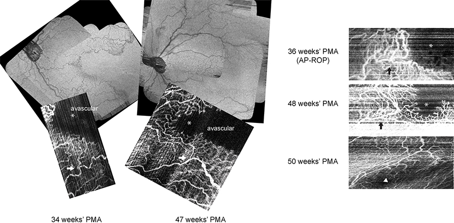
Aggressive posterior retinopathy of prematurity (AP-ROP) is a severe form of ROP occurring in preterm infants that is characterized by rapid progression and prominent vascularity. We report the use of investigational bedside noninvasive optical coherence tomography angiography to visualize the slow and progressive perifoveal vascular formation in an infant with AP-ROP treated with bevacizumab. We also document extensive vascular shunts and morphological differences between arrested and growing retinal capillaries at the vascular wavefront.
Trial registration: ClinicalTrials.gov NCT02887157.
Copyright © 2020 American Association for Pediatric Ophthalmology and Strabismus. Published by Elsevier Inc. All rights reserved.
Figures
References
-
- International Committee for the Classification of Retinopathy of Prematurity. The International Classification of Retinopathy of Prematurity revisited. Arch Ophthalmol 2005;123:991–9. - PubMed
-
- Lepore D, Quinn GE, Molle F, et al. Intravitreal bevacizumab versus laser treatment in type 1 retinopathy of prematurity: report on fluorescein angiographic findings. Ophthalmology 2014;121:2212–19. - PubMed
-
- Lorenz B, Stieger K, Jager M, Mais C, Stieger S, Andrassi-Darida M. Retinal vascular development with 0.312 mg intravitreal bevacizumab to treat severe posterior retinopathy of prematurity: a longitudinal fluorescein angiographic study. Retina 2017;37:97–111. - PubMed
-
- Lepore D, Ji MH, Quinn GE, et al. Functional and morphologic findings at four years after intravitreal bevacizumab or laser for type 1 ROP. Ophthalmic Surg Lasers Imaging Retina 2020;51:180–86. - PubMed
Publication types
MeSH terms
Substances
Associated data
Grants and funding
LinkOut - more resources
Full Text Sources
Medical
