. 2020 Oct:37:101747.
doi: 10.1016/j.redox.2020.101747. Epub 2020 Oct 7.

Meteorin-like protein attenuates doxorubicin-induced cardiotoxicity via activating cAMP/PKA/SIRT1 pathway

Affiliations

Meteorin-like protein attenuates doxorubicin-induced cardiotoxicity via activating cAMP/PKA/SIRT1 pathway

Can Hu et al. Redox Biol. 2020 Oct.

Abstract

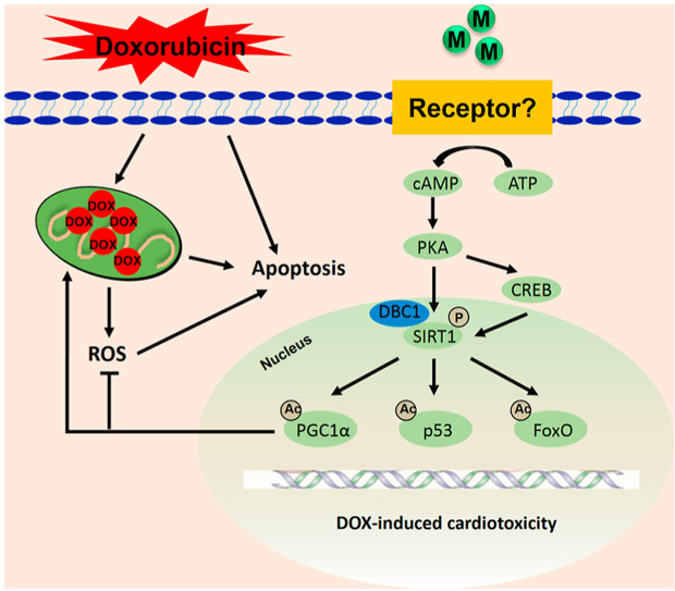
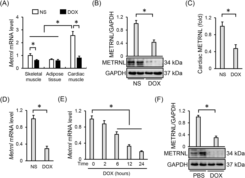
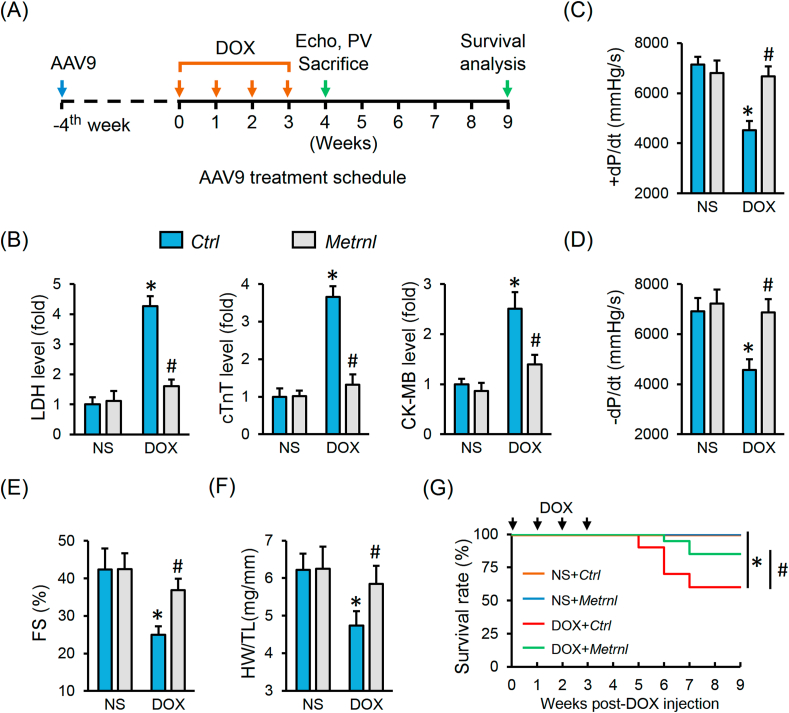
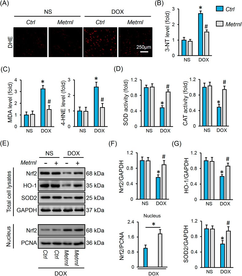
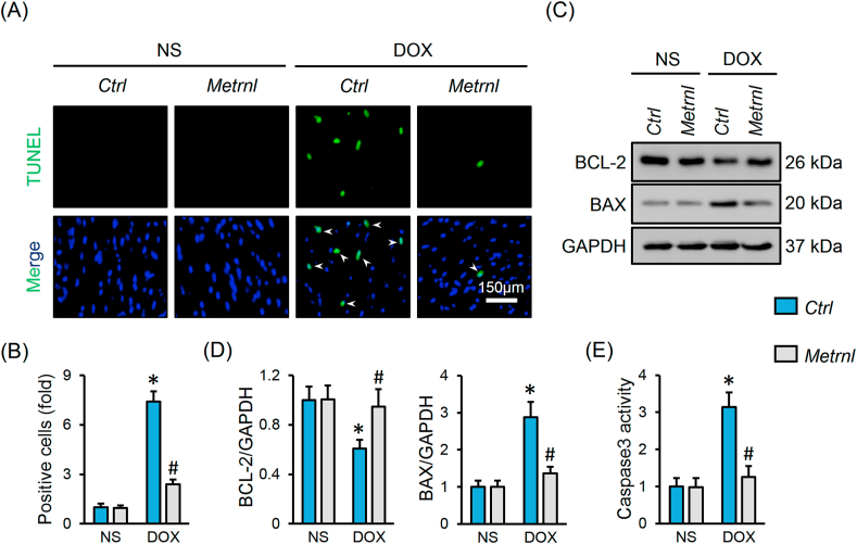
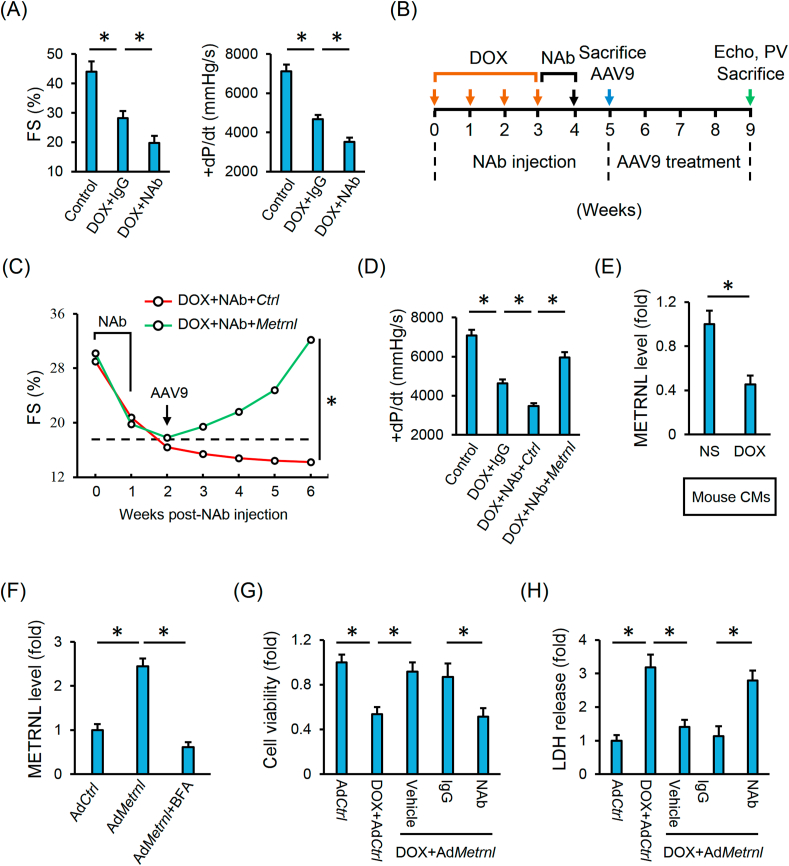
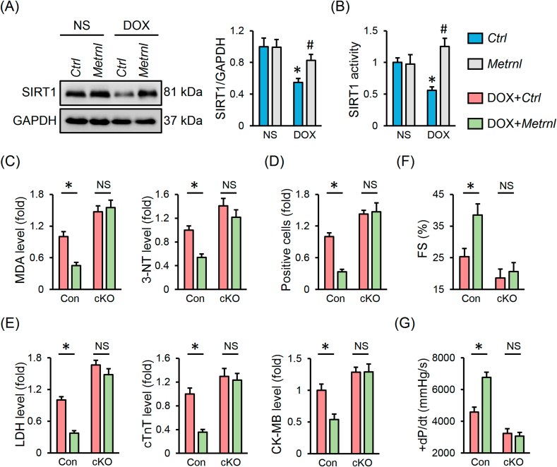
Meteorin-like (METRNL) protein is a newly identified myokine that functions to modulate energy expenditure and inflammation in adipose tissue. Herein, we aim to investigate the potential role and molecular basis of METRNL in doxorubicin (DOX)-induced cardiotoxicity. METRNL was found to be abundantly expressed in cardiac muscle under physiological conditions that was decreased upon DOX exposure. Cardiac-specific overexpression of METRNL by adeno-associated virus serotype 9 markedly improved oxidative stress, apoptosis, cardiac dysfunction and survival status in DOX-treated mice. Conversely, knocking down endogenous METRNL by an intramyocardial injection of adenovirus exacerbated DOX-induced cardiotoxicity and death. Meanwhile, METRNL overexpression attenuated, while METRNL silence promoted oxidative damage and apoptosis in DOX-treated H9C2 cells. Systemic METRNL depletion by a neutralizing antibody aggravated DOX-related cardiac injury and dysfunction in vivo, which were notably alleviated by METRNL overexpression within the cardiomyocytes. Besides, we detected robust METRNL secretion from isolated rodent hearts and cardiomyocytes, but to a less extent in those with DOX treatment. And the beneficial effects of METRNL in H9C2 cells disappeared after the incubation with a METRNL neutralizing antibody. Mechanistically, METRNL activated SIRT1 via the cAMP/PKA pathway, and its antioxidant and antiapoptotic capacities were blocked by SIRT1 deficiency. More importantly, METRNL did not affect the tumor-killing action of DOX in 4T1 breast cancer cells and tumor-bearing mice. Collectively, cardiac-derived METRNL activates SIRT1 via cAMP/PKA signaling axis in an autocrine manner, which ultimately improves DOX-elicited oxidative stress, apoptosis and cardiac dysfunction. Targeting METRNL may provide a novel therapeutic strategy for the prevention of DOX-associated cardiotoxicity.

Keywords: Apoptosis; Doxorubicin; METRNL; Oxidative stress; SIRT1.


Conflict of interest statement

None declared.

Figures

Image 1
Graphical abstract
Fig. 1
Fig. 1
METRNL is downregulated in hearts and cardiomyocytes upon DOX stimulation. (A) Relative mRNA levels of METRNL in different mouse tissues under DOX treatment (n = 6). (B–C) Cardiac METRNL expression detected by Western blot and ELISA (n = 6). (D) Relative Metrnl mRNA level in the adult mouse cardiomyocytes isolated from the hearts with or without DOX treatment (n = 6). (E) Relative Metrnl mRNA level in H9C2 cells under DOX stimulation with different times (n = 6). (F) METRNL protein alteration in H9C2 cells after DOX incubation for 24 h (n = 6). Values represent the mean ± SD. *P < 0.05 versus the matched group.
Fig. 2
Fig. 2
METRNL overexpression alleviates DOX-induced cardiotoxicity in mice. (A) Schematic protocol for AAV9 and DOX treatment. (B) Serum levels of LDH, cTnT and CK-MB in mice with or without METRNL overexpression after DOX injection (n = 8). (CE) Hemodynamic and echocardiographic parameters of cardiac function (n = 8). (F) Statistical data of HW/TL (n = 8). (G) The survival curves up to 9 weeks after the first DOX injection in mice with or without METRNL overexpression (n = 20). Values represent the mean ± SD. *P < 0.05 versus NS + Ctrl, #P < 0.05 versus DOX + Ctrl.
Fig. 3
Fig. 3
METRNL overexpression inhibits oxidative stress in DOX-treated hearts. (A) Representative images of DHE staining in heart samples (n = 6). (B–C) The levels of 3-NT, MDA and 4-HNE in heart samples (n = 6–8). (D) Total SOD and CAT activities in hearts (n = 8). (EG) Representative Western blot images and statistical results (n = 6). Values represent the mean ± SD. *P < 0.05 versus NS + Ctrl, #P < 0.05 versus DOX + Ctrl. In Fig. 3F, *P < 0.05 versus the matched group.
Fig. 4
Fig. 4
METRNL overexpression inhibits apoptosis in DOX-treated hearts. (AB) Representative images of TUNEL staining and the quantitative data about TUNEL-positive nuclei in heart samples (n = 8). White arrows indicate TUNEL-positive nuclei. (CD) Representative Western blot images and statistical results (n = 6). (E) Caspase3 activity in heart samples (n = 8). Values represent the mean ± SD. *P < 0.05 versus NS + Ctrl, #P < 0.05 versus DOX + Ctrl.
Fig. 5
Fig. 5
Cardiac-derived METRNL prevents DOX-induced cardiotoxicity in an autocrine manner. (A) Hemodynamic and echocardiographic parameters of cardiac function in mice with or without NAb injection (n = 5). (B) Schematic protocol for NAb, DOX and AAV9 treatment. (CD) Hemodynamic and echocardiographic parameters of cardiac function (n = 5). (E) Adult mouse CMs were isolated from the mice with or without DOX injection and then incubated in serum-free medium for additional 4 h to detect METRNL secretion (n = 6). (F) H9C2 cells were infected with the adenovirus and cultured in normal medium for 24 h to allow gene manipulation, which were then treated with BFA (2.5 μmol/L) in serum-free medium for additional 4 h to detect METRNL secretion (n = 6). (G) Cell viability analysis by CCK-8 method (n = 8). (H) LDH release in H9C2 cells (n = 6). Values represent the mean ± SD. *P < 0.05 versus the matched group.
Fig. 6
Fig. 6
METRNL exerts the cardioprotective effects via activating SIRT1 in vivo. (A) Cardiac SIRT1 protein abundance and statistical results (n = 6). (B) Cardiac SIRT1 activity (n = 8). (C) The levels of MDA and 3-NT in heart samples (n = 6–8). (D) Quantification of RUNEL-positive nuclei in heart tissues (n = 8). (E) Serum levels of LDH, cTnT and CK-MB in mice (n = 8). (FG) Hemodynamic and echocardiographic parameters (n = 8). Values represent the mean ± SD. *P < 0.05 versus the matched group. In Fig. 6A and B, *P < 0.05 versus NS + Ctrl, #P < 0.05 versus DOX + Ctrl. NS indicates no significance.

References

    1. Armenian S.H., Lacchetti C., Barac A., Carver J., Constine L.S., Denduluri N. Prevention and monitoring of cardiac dysfunction in survivors of adult cancers: American society of clinical oncology clinical practice guideline. J. Clin. Oncol. 2017;35:893–911. - PubMed
    1. Wallace K.B., Sardao V.A., Oliveira P.J. Mitochondrial determinants of doxorubicin-induced cardiomyopathy. Circ. Res. 2020;126:926–941. - PMC - PubMed
    1. Doroshow J.H., Locker G.Y., Myers C.E. Enzymatic defenses of the mouse heart against reactive oxygen metabolites: alterations produced by doxorubicin. J. Clin. Invest. 1980;65:128–135. - PMC - PubMed
    1. Zhang X., Hu C., Kong C.Y., Song P., Wu H.M., Xu S.C. FNDC5 alleviates oxidative stress and cardiomyocyte apoptosis in doxorubicin-induced cardiotoxicity via activating AKT. Cell Death Differ. 2020;27:540–555. - PMC - PubMed
    1. Hu C., Zhang X., Wei W., Zhang N., Wu H., Ma Z. Matrine attenuates oxidative stress and cardiomyocyte apoptosis in doxorubicin-induced cardiotoxicity via maintaining AMPKalpha/UCP2 pathway. Acta Pharm. Sin. B. 2019;9:690–701. - PMC - PubMed

Publication types

LinkOut - more resources