Review
. 2021 Mar;46(3):200-212.
doi: 10.1016/j.tibs.2020.09.006. Epub 2020 Oct 9.

Balancing the Opposing Principles That Govern Peroxisome Homeostasis

Affiliations
Review

Balancing the Opposing Principles That Govern Peroxisome Homeostasis

Shanmuga S Mahalingam et al. Trends Biochem Sci. 2021 Mar.

Abstract

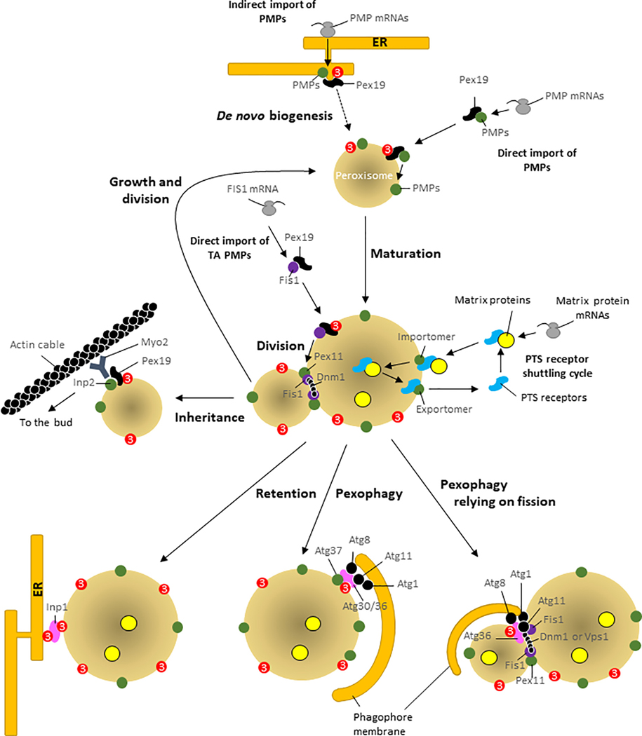
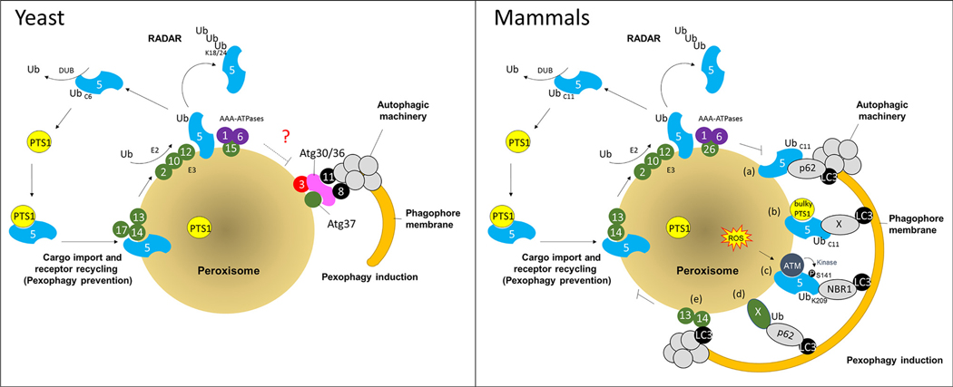
Despite major advances in our understanding of players and mechanisms involved in peroxisome biogenesis and peroxisome degradation, very few studies have focused on unraveling the multi-layered connections between, and the coordination of, these two opposing processes that regulate peroxisome homeostasis. The intersection between these processes also provides exciting avenues for future research. This review highlights the links between peroxisome biogenesis and degradation, incorporating an integrative approach that is critical not only for a mechanistic understanding, but also for manipulating the balance between these processes in relevant disease models.

Keywords: crosstalk; homeostasis; peroxisome biogenesis; peroxisome disorders; pexophagy.


Figures

Figure 1.
Figure 1.. Peroxisome Homeostasis and Key Roles of Pex3 in the Fate of Peroxisomes.
Direct and indirect import of PMPs: direct PMP import to the membrane requires Pex19, which docks with Pex3 and inserts PMPs into the membrane (growth and division model). Alternatively, PMPs may be imported indirectly via the ER, wherein PMPs bud from the ER using Pex3 and Pex19, and the resulting vesicles fuse with pre-existing peroxisomes. In cells lacking peroxisomes, the vesicles containing PMPs fuse and grow to form new peroxisomes (de novo biogenesis model). Direct import of TA proteins: these (e.g., Fis1) are imported post-translationally to the peroxisome membrane using Pex19 and Pex3. Maturation and PTS receptor shuttling cycle: proteins destined for the peroxisomal matrix are translocated by PTS receptors across the importomer. Following cargo release in the matrix, PTS receptors recycle back to the cytosol, via the exportomer. Division: another common mode of peroxisome proliferation is by growth and division of pre-existing peroxisomes, during which daughter peroxisomes bud from a mother peroxisome, using the division machinery comprised of Pex11, Fis1, and Dnm1 in yeast. Inheritance and Retention: during cytokinesis in yeast, daughter peroxisomes are inherited using the Myo2 motor interacting with peroxisomal Inp2 and actin filaments, whereas the peroxisome in the mother cell is retained by the interaction of Inp1 with Pex3 at the cortical ER. Pexophagy: requires SARs (Atg30, Atg36) and a SAR regulator (Atg37 in P. pastoris), which associate with, or are inserted into, the peroxisome membrane by Pex3. SARs activated by environmental cues engage the autophagy machinery (Atg8, Atg11, and Atg1 kinase) allowing phagophore membranes to engulf peroxisomes. Pexophagy relying on fission: in S. cerevisiae, pexophagy requires peroxisome division. Abbreviations: ER, endoplasmic reticulum; PMPs, peroxisomal membrane proteins; PTS, peroxisomal targeting signal; TA, tail-anchored; SARs, selective autophagy receptors.
Figure 2.
Figure 2.. Role of Pex5 in Biogenesis and Pexophagy in Yeast and Mammalian Cells.
During peroxisome biogenesis in both yeast (left panel) and mammals (right panel) Pex5 recognizes the PTS1-containing cargoes, and transports them into the peroxisome matrix after interacting with the importomer proteins (conserved components are Pex13 and Pex14). After cargo release, Pex5 recycles back to the cytoplasm using exportomer components, Pex1 and Pex6, anchored at the peroxisome membrane through Pex15 in yeast and PEX26 in mammals. Receptor recycling requires Pex5 mono-ubiquitylation (at residue C6 in yeast and C11 in mammals), via an E2 enzyme (Pex4 in yeast and UbcH5 in mammals) and RING E3 ligases (Pex2, Pex10, and Pex12). If Pex5 recycling is blocked, then it can be polyubiquitylated and targeted for UPS-mediated turnover (RADAR pathway). Alternatively, when peroxisomes are damaged or redundant, pexophagy is activated. In yeast, a block in receptor recycling triggers pexophagy. It is still unclear (marked as ‘?’) how the yeast the AAA-ATPases regulate pexophagy. In mammals, either (a) the accumulation of mono-ubiquitylated PEX5 at the peroxisome membrane, or (b) the presence of a bulky PTS1 that cannot be released form PEX5, can activate pexophagy [24]. Alternatively (c), as a part of quality control, under conditions of oxidative stress, the ATM kinase phosphorylates PEX5 at S141, leading to its subsequent ubiquitylation by the RING E3 complex at K209, and activation of pexophagy [25]. Pexophagy can also be triggered in mammals in a PEX5-independent manner, either (d) by ubiquitination of PMPs (marked by ‘X’), such as PEX3 and PMP70, which are then recognized by autophagy adaptor proteins NBR1, and/or p62 [27], or (e) by direct binding of PEX14 to LC3 [26]. Abbreviations: ATM, ataxia-telangiectasia mutated; DUB, deubiquitylating enzyme; PTS1, peroxisomal targeting signal 1; RADAR, receptor accumulation and degradation in the absence of recycling; Ub, ubiquitin; UPS, ubiquitin-proteasome system; ROS, reactive oxygen species.
Figure 3.
Figure 3.. Peroxisome and Pexophagosome Movement Along the Cytoskeleton in Yeast and Mammals.
In yeast (left panel), newly-divided peroxisomes containing Inp2, interacting with Pex19 (which is bound to Pex3), engage the Myo2 motor on actin filaments to move peroxisomes to the bud. During pexophagy, the phagophore membrane expands by the delivery of vesicles to the PAS (site of initiation of pexophagy) using the actin cytoskeleton. The resulting pexophagosome moves to the vacuole along the actin cytoskeleton using unknown receptors on the pexophagosome. In mammals (right panel), peroxisomes move using kinesin or dynein motors, in the anterograde (cell periphery) and retrograde (cell interior) directions, respectively. These motors engage with microtubules. Phagophore formation relies on the delivery of membrane vesicles from different sources to the omegasome (site of initiation of pexophagy), using the Myosin II motor moving along the actin cytoskeleton network. Pexophagy uses PEX5-dependent or -independent SARs on peroxisomes to engage selective autophagy adaptors (NBR1 or p62). The pexophagosome resulting from peroxisome engulfment by the phagophore membrane also uses kinesin and dynein motors moving along microtubules. Abbreviations: PAS, pre-autophagosomal structure; PMPs, peroxisomal membrane proteins; SARs, selective autophagy receptors.

References

    1. Argyriou C. et al. (2016) Peroxisome biogenesis disorders. Transl. Sci. Rare Dis. 1, 111–144 - PMC - PubMed
    1. Agrawal G and Subramani S. (2016) De novo peroxisome biogenesis: evolving concepts and conundrums. Biochim. Biophys. Acta 1863, 892–901 - PMC - PubMed
    1. Farre JC et al. (2019) Peroxisome biogenesis, membrane contact sites, and quality control. EMBO Rep. 20, e46864 - PMC - PubMed
    1. Hettema EH et al. (2014) Evolving models for peroxisome biogenesis. Curr. Opin. Cell Biol. 29, 25–30 - PMC - PubMed
    1. Meinecke M et al. (2010) The peroxisomal importomer constitutes a large and highly dynamic pore. Nat. Cell Biol. 12, 273–277 - PubMed

Publication types

MeSH terms