. 2020 Dec;216(4):1103-1116.
doi: 10.1534/genetics.120.303623. Epub 2020 Oct 12.

Plasma Membrane Fusion Is Specifically Impacted by the Molecular Structure of Membrane Sterols During Vegetative Development of Neurospora crassa

Affiliations

Plasma Membrane Fusion Is Specifically Impacted by the Molecular Structure of Membrane Sterols During Vegetative Development of Neurospora crassa

Martin Weichert et al. Genetics. 2020 Dec.

Abstract

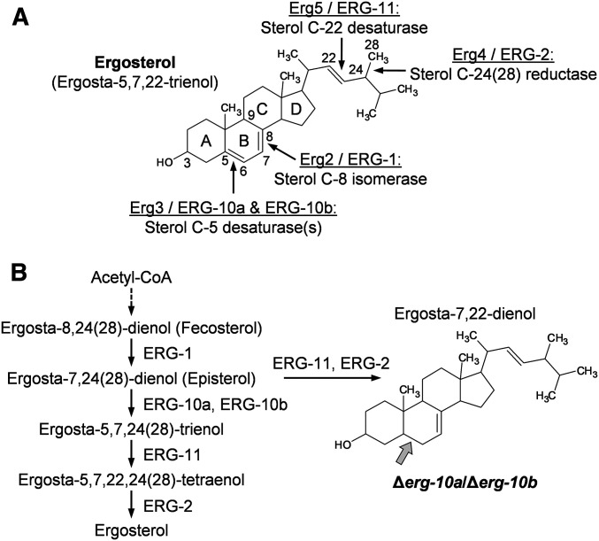
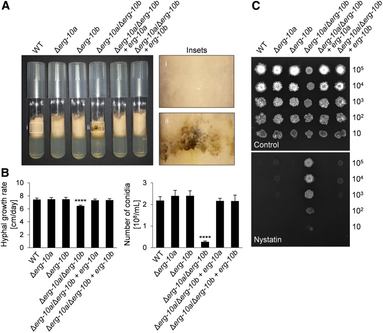
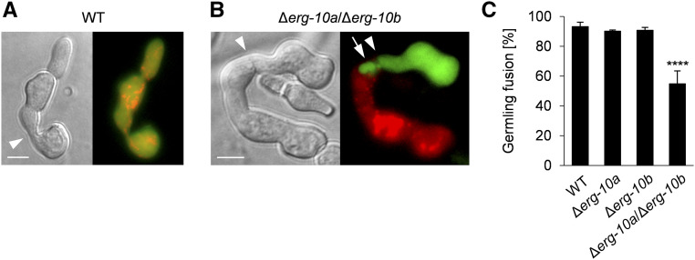
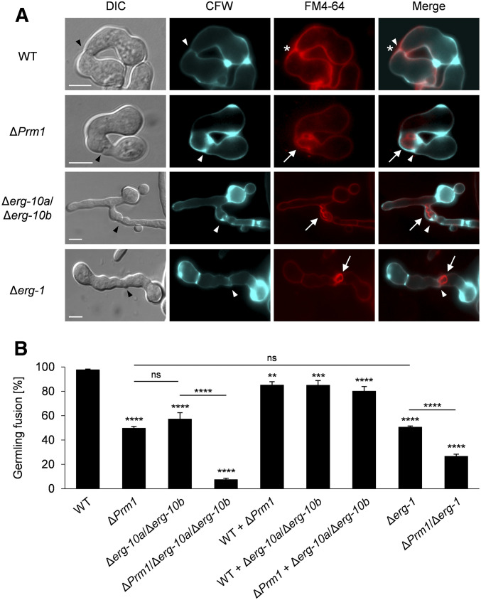
Cell-to-cell fusion is crucial for the development and propagation of most eukaryotic organisms. Despite this importance, the molecular mechanisms mediating this process are only poorly understood in biological systems. In particular, the step of plasma membrane merger and the contributing proteins and physicochemical factors remain mostly unknown. Earlier studies provided the first evidence of a role of membrane sterols in cell-to-cell fusion. By characterizing different ergosterol biosynthesis mutants of the fungus Neurospora crassa, which accumulate different ergosterol precursors, we show that the structure of the sterol ring system specifically affects plasma membrane merger during the fusion of vegetative spore germlings. Genetic analyses pinpoint this defect to an event prior to engagement of the fusion machinery. Strikingly, this effect is not observed during sexual fusion, suggesting that the specific sterol precursors do not generally block membrane merger, but rather impair subcellular processes exclusively mediating fusion of vegetative cells. At a colony-wide level, the altered structure of the sterol ring system affects a subset of differentiation processes, including vegetative sporulation and steps before and after fertilization during sexual propagation. Together, these observations corroborate the notion that the accumulation of particular sterol precursors has very specific effects on defined cellular processes rather than nonspecifically disturbing membrane functioning. Given the phenotypic similarities of the ergosterol biosynthesis mutants of N. crassa during vegetative fusion and of Saccharomyces cerevisiae cells undergoing mating, our data support the idea that yeast mating is evolutionarily and mechanistically more closely related to vegetative than sexual fusion of filamentous fungi.

Keywords: Neurospora crassa; cell fusion; ergosterol; mating; plasma membrane fusion.


Figures

Figure 1
Figure 1
The sterol C-5 desaturases ERG-10a and ERG-10b have a redundant function in ergosterol biosynthesis in N. crassa. (A) Structure of ergosterol. Enzymes and their functions mediating double-bond arrangements in ergosterol precursors are depicted (S. cerevisiae/N. crassa). Numbers along ergosterol denote the position of selected carbon atoms, and letters indicate the carbon rings. (B) Late steps in the hypothetical biosynthesis pathway of ergosterol in N. crassa showing the reactions catalyzed by the enzymes presented in (A). ERG-10a and ERG-10b mediate the same reaction, whose block results in the accumulation of a sterol intermediate with an altered ring structure (gray arrow).
Figure 2
Figure 2
Impact of Δerg-10a and Δerg-10b on vegetative growth of N. crassa. (A) Growth of WT (FGSC 2489), Δerg-10a (FGSC 20057), Δerg-10b (N4-30), Δerg-10aerg-10b (N4-38), Δerg-10aerg-10b + erg-10a (MW_630), and Δerg-10aerg-10b + erg-10b (MW_631) in MM tubes. Insets: in contrast to WT (i), Δerg-10aerg-10b (ii) typically produces dark pigments along the glass wall of the tube. (B) Quantification of linear hyphal extension rates and sporulation. Values represent the mean ± SD from at least three independent experiments per strain (****P < 0.0001, ANOVA with Tukey’s post hoc test). (C) Ten-fold serial dilutions of conidia of the indicated strains were spotted onto sorbose-rich medium in the absence or presence of the polyene antifungal compound nystatin. FGSC, Fungal Genetics Stock Center; MM, minimal medium; WT, wild-type.
Figure 3
Figure 3
Germlings of ∆erg-10a/erg-10b are impaired in cell-to-cell fusion. (A) Microscopic analysis of cell-to-cell fusion in a pair of WT germlings expressing cytosolic GFP and Cherry Red (N3-06 + N3-07) after physical contact (arrowhead), resulting in mixing of green and red fluorescence. (B) A pair of Δerg-10aerg-10b germlings expressing cytosolic GFP or Cherry Red (MW_175 + MW_179). Note the lack of cytoplasmic mixing and the extrusion extending from one cell into the other (arrow). Bar in (A and B), 5 μm. (C) Quantification of germling fusion in cell pairings of WT (N3-06 + N3-07), Δerg-10a (N5-01 + N5-02), Δerg-10b (MW_103 + MW_105), and Δerg-10aerg-10b (MW_175 + MW_179). Values represent the mean ± SD from at least three independent cell populations with 87–109 germling pairs per replicate (****P < 0.0001, ANOVA with Tukey’s post hoc test). WT, wild-type.
Figure 4
Figure 4
Germlings of the mutants Δerg-10a Δerg-10b and Δerg-1 are defective in plasma membrane merger during cell-to-cell fusion. (A) Costaining with CFW and FM4-64 to visualize the cell wall and the plasma membrane, respectively. Black arrowheads indicate cell contacts. Note that WT (FGSC 2489) pairings form both an opening in the cell wall (white arrowhead) and a fusion pore in the membrane (asterisk). The mutants ΔPrm1 (A32), Δerg-10aerg-10b (N4-38), and Δerg-1 (MW_308) form membrane invaginations (arrows) that stretch through the cell wall opening from one into the other cell (white arrowheads). Bar, 5 μm. (B) Quantification of germling fusion of erg mutants carrying the additional deletion of Prm1. Values represent the mean ± SD from three independent experiments per set of strains with a range of 40–211 germling pairs per population (**P < 0.01, ***P < 0.001, ****P < 0.0001; ANOVA with Tukey’s post hoc test). See Figure S2 for images of representative germling pairs from these experiments. CFW, calcofluor white; ns, not significant; WT, wild-type.
Figure 5
Figure 5
The various plasma membrane fusion mutants differ in the frequency of fusion-induced cell lysis. Quantitative analysis of cell lysis in germling pairs of WT (FGSC 2489) and mutants lacking erg genes and/or Prm1 on MM, with normal and reduced levels of Ca2+. Values represent the mean ± SD from three independent populations of 100–125 germling pairs per replicate, strain, and growth condition. Statistical analysis with ANOVA and Tukey’s post hoc test: **P < 0.01, ***P < 0.001, ****P < 0.0001 for comparisons with ΔPrm1 at normal Ca2+ concentration; horizontal lines denote additional comparisons between strains (***P < 0.001); # denotes a significant difference (P < 0.001) for comparisons of ΔPrm1 between normal and half Ca2+ concentration, with no significant differences for any of the other strains between these two growth conditions. FGSC, Fungal Genetics Stock Center; MM, minimal medium; ns, not significant; WT, wild-type.
Figure 6
Figure 6
The deletion of erg-10a/erg-10b does not impact mating fusion in N. crassa. (A) Representative images of mating pairs in homozygous crossings of WT (FGSC 988 × N2-12) and Δerg-10aerg-10b (N4-37 × MW_181). Microconidia (black arrows) expressing H1-GFP were observed before (24 hr) and after (48 hr) contact with a trichogyne of the opposite mating type (mat a × mat A). The disappearance of the nuclear fluorescence in the spores (white arrows) indicates cell-to-cell fusion events, resulting in the transition of nuclei via the trichogynes into the fruiting bodies. Bar, 10 μm. (B) Quantification of fusion between trichogynes and microconidia. Values represent the mean ± SD from three to six independent crossings for each combination with 39–122 mating pairs per replicate (no statistically significant differences were detected between these crosses: P > > 0.05, ANOVA with Dunnett’s post hoc test). FGSC, Fungal Genetics Stock Center; WT, wild-type.
Figure 7
Figure 7
Membrane sterol distribution differs between vegetative and sexual cells of N. crassa. Germlings and trichogynes were stained with filipin. (A) Accumulation of sterols at the polarized cell tips of WT (FGSC 2489), Δerg-10aerg-10b (N4-38), and Δerg-1 (MW_308) germlings. (B) Arrows indicate sterol-rich domains in interacting WT germlings (g). The white square marks the area of the enlarged image to the right. (C) Trichogyne (t)–microconidium (m) interaction. The white rectangle indicates the position of the enlarged image to the right. Bar in (A–C), 5 μm. (D) Quantification of sterol-rich cell tips in germlings and trichogynes during cell-to-cell interactions. Values represent the mean ± SD from three and two independent experiments with ∼100 germling pairs and up to 40 trichogynes per replicate (****P < 0.0001, unpaired, two-tailed Student’s t-test). FGSC, Fungal Genetics Stock Center; WT, wild-type.
Figure 8
Figure 8
Fertility of the Δerg-10aerg-10b mutant is affected by pre- and postfertilization deficiencies. (A) Fruiting body development in homozygous and heterozygous crosses (mat A × mat a) of WT (FGSC 2489 and FGSC 988) and Δerg-10aerg-10b (N4-38 and N4-37). (B) Microscopic analysis of rosettes of asci isolated from the fruiting bodies shown in (A). Inset images show the amount of ascospores harvested from one representative plate per crossing. (C and D) Analysis of perithecia, asci, and ascospores (insets) from crosses with a strain carrying the suppressor mutation ΔSad-1 (FGSC 11151) and/or involving the helper strain (FGSC 4564). Note that only the presence of the helper and the ΔSad-1 mutation fully restore fertility of the crosses. Bars in A and C, 500 μm; B and D, 200 μm. FGSC, Fungal Genetics Stock Center; WT, wild-type.

Similar articles

Cited by

References

    1. Aguilar P. S., Engel A., and Walter P., 2007. The plasma membrane proteins Prm1 and Fig1 ascertain fidelity of membrane fusion during yeast mating. Mol. Biol. Cell 18: 547–556. 10.1091/mbc.e06-09-0776 - DOI - PMC - PubMed
    1. Aguilar P. S., Heiman M. G., Walther T. C., Engel A., Schwudke D. et al. , 2010. Structure of sterol aliphatic chains affects yeast cell shape and cell fusion during mating. Proc. Natl. Acad. Sci. USA 107: 4170–4175. 10.1073/pnas.0914094107 - DOI - PMC - PubMed
    1. Aguilar P. S., Baylies M. K., Fleissner A., Helming L., Inoue N. et al. , 2013. Genetic basis of cell-cell fusion mechanisms. Trends Genet. 29: 427–437. 10.1016/j.tig.2013.01.011 - DOI - PMC - PubMed
    1. Athanasopoulos A., André B., Sophianopoulou V., and Gournas C., 2019. Fungal plasma membrane domains. FEMS Microbiol. Rev. 43: 642–673. doi: 10.1093/femsre/fuz022 - PubMed
    1. Bistis G. N., 1981. Chemotropic interactions between trichogynes and conidia of opposite mating-type in Neurospora crassa. Mycologia 73: 959–975. 10.1080/00275514.1981.12021425 - DOI

Publication types

MeSH terms