. 2020 Oct 27;117(43):26915-26925.
doi: 10.1073/pnas.2001046117. Epub 2020 Oct 12.

Swine acute diarrhea syndrome coronavirus replication in primary human cells reveals potential susceptibility to infection

Affiliations

Swine acute diarrhea syndrome coronavirus replication in primary human cells reveals potential susceptibility to infection

Caitlin E Edwards et al. Proc Natl Acad Sci U S A. .

Abstract

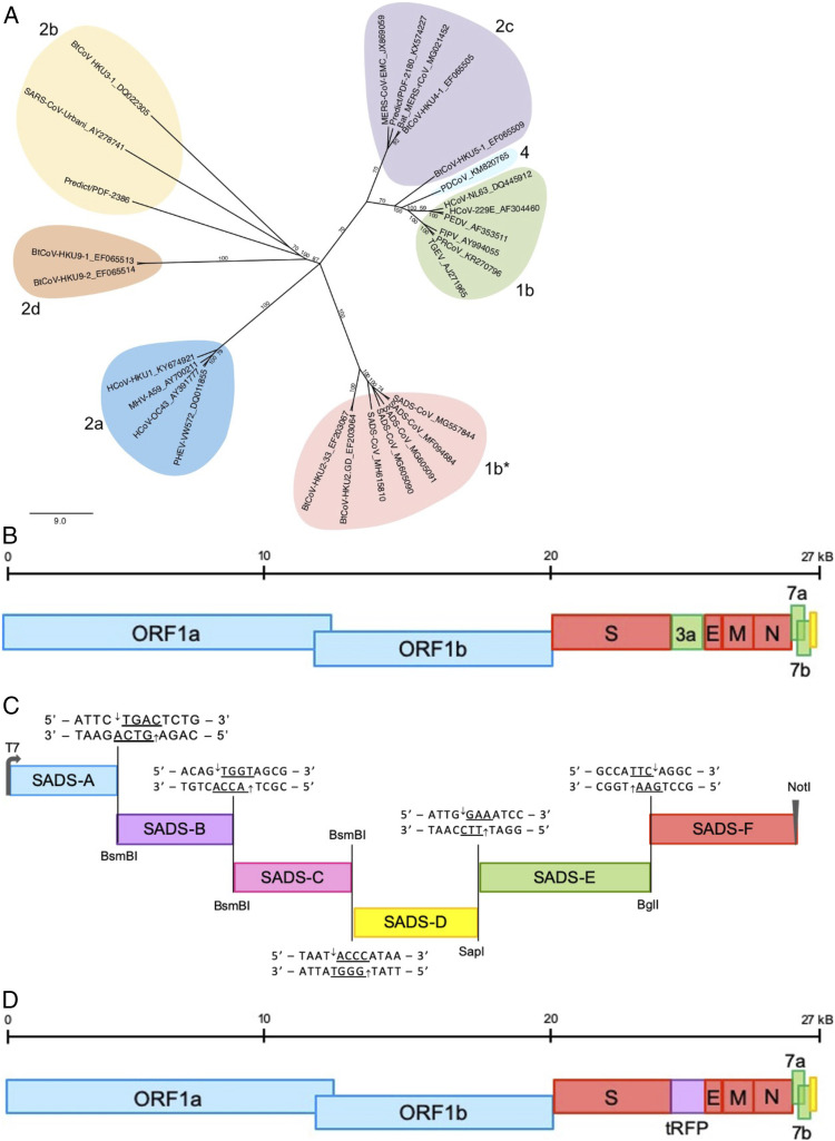
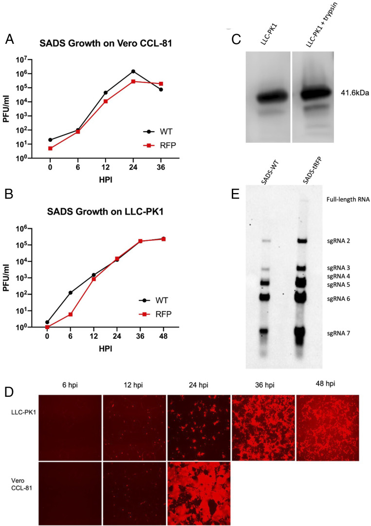
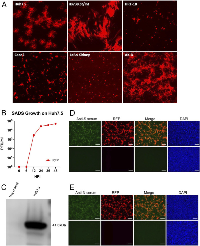
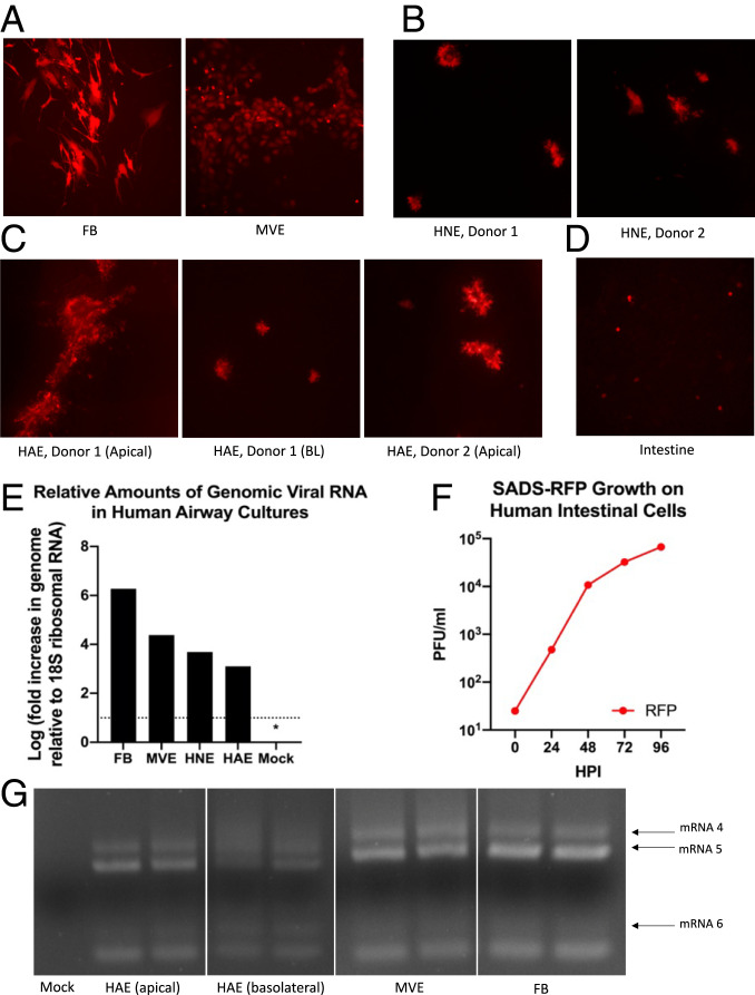
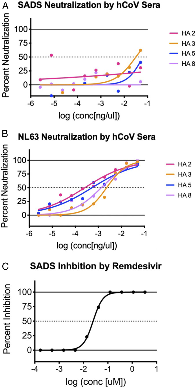
Zoonotic coronaviruses represent an ongoing threat, yet the myriads of circulating animal viruses complicate the identification of higher-risk isolates that threaten human health. Swine acute diarrhea syndrome coronavirus (SADS-CoV) is a newly discovered, highly pathogenic virus that likely evolved from closely related HKU2 bat coronaviruses, circulating in Rhinolophus spp. bats in China and elsewhere. As coronaviruses cause severe economic losses in the pork industry and swine are key intermediate hosts of human disease outbreaks, we synthetically resurrected a recombinant virus (rSADS-CoV) as well as a derivative encoding tomato red fluorescent protein (tRFP) in place of ORF3. rSADS-CoV replicated efficiently in a variety of continuous animal and primate cell lines, including human liver and rectal carcinoma cell lines. Of concern, rSADS-CoV also replicated efficiently in several different primary human lung cell types, as well as primary human intestinal cells. rSADS-CoV did not use human coronavirus ACE-2, DPP4, or CD13 receptors for docking and entry. Contemporary human donor sera neutralized the group I human coronavirus NL63, but not rSADS-CoV, suggesting limited human group I coronavirus cross protective herd immunity. Importantly, remdesivir, a broad-spectrum nucleoside analog that is effective against other group 1 and 2 coronaviruses, efficiently blocked rSADS-CoV replication in vitro. rSADS-CoV demonstrated little, if any, replicative capacity in either immune-competent or immunodeficient mice, indicating a critical need for improved animal models. Efficient growth in primary human lung and intestinal cells implicate SADS-CoV as a potential higher-risk emerging coronavirus pathogen that could negatively impact the global economy and human health.

Keywords: One Health; SADS; coronavirus; emerging infectious disease.


Conflict of interest statement

The authors declare no competing interest.

Figures

Fig. 1.
Fig. 1.
Spike phylogeny of representative coronaviruses and organization of the SADS-CoV wild-type and RFP infectious clones. (A) The S protein sequences of selected coronaviruses were aligned and phylogenetically compared. Coronavirus genera are grouped by classic subgroup designations (1b, 2a–d, 4). Sequences designated as 1b* (including SADS-CoV and related viruses) group with 1b viruses in proteins other than S. Sequences were aligned using free end gaps with the Blosum62 cost matrix in Geneious Prime. The tree was constructed using the neighbor-joining method based on the multiple sequence alignment, also in Geneious Prime. Numbers following the underscores in each sequence correspond to the GenBank accession number. The radial phylogram was rendered for publication using Adobe Illustrator CC 2019. (B) The general arrangement of the SADS-CoV genome. Blue represents nonstructural proteins ORF1a and ORF1b. Red represents structural proteins spike, envelope, membrane, and nucleocapsid. Green represents accessory proteins 3a, 7a, and 7b. Yellow represents the untranslated region. (C) The full-length infectious clone was divided into six contiguous cDNAs flanked by either BsmBI (SADS-CoV A–C), SapI (SADS-CoV D), BglI (SADS-CoV E–F) to allow for efficient assembly of the full-length SADS cDNA. BsmBI and SapI are not present in the viral genome sequence but are introduced externally in the fragment plasmid sequence. SADS-CoV A (nucleotides 1 to 4,496), SADS-CoV B (nucleotides 4,497 to 8,996), SADS-CoV C (nucleotides 8,997 to 13,496), SADS-CoV D (nucleotides 13,497 to 17,997), SADS-CoV E (nucleotides 17,998 to 22,892), and SADS-CoV F (nucleotides 22,893 to end). (D) General organization of SADS-CoV tRFP depicting the insertion of tRFP in place of NS3a.
Fig. 2.
Fig. 2.
Growth of wild-type and tRFP SADS-CoV. Growth of SADS-CoV wild-type and tRFP infectious clones compared in Vero CCL-81 (A) and LLC-PK1 (B) cultures. All supernatants were titered on Vero CCL-81 cells. Viral titers grew to mid-105 to 107 PFU/mL in these cell types, with the highest titers seen in Vero CCL-81 cells. (C) Cultures of LLC-PK1 cells infected with SADS-CoV and protein lysates were run on Western blots with and without trypsin for comparison. Cell lysates were probed with antinucleocapsid antibodies from sera of mice immunized with VRP. N proteins (41.6 kDa) were present among both conditions. (D) tRFP expression of Vero CCL-81 and LLC-PK1 cultures is shown at 10× magnification. By 36 h, cytopathic effect in Vero CCL-81 cultures was complete. (E) Northern blot analysis of wild-type SADS and SADS tRFP for leader containing transcripts of known ORFs.
Fig. 3.
Fig. 3.
Host range of SADS tRFP. (A) Cultures of human Huh7.5 liver, human Hs738.St/Int stomach-intestine, human colo-rectal HRT-18 tumor, Caco2, LaBo kidney, and feline AK-D lung cells were infected with SADS tRFP. All cell types, with the exception of Caco2 and Huh7.5 cells, were cultured in the presence of trypsin to enhance virus infectivity. Cultures were visualized at 48 hpi for fluorescence at 10× magnification. (B) Growth of SADS-tRFP in Huh 7.5 cells. (C) Cultures of Huh7.5 cells were infected with SADS-CoV and lysed for analysis by Western blot. N protein has a molecular mass of ∼41.6 kDa. Immunofluorescent images of Vero CCL-81 cells infected with mock or SADS-CoV tRFP virus and stained with mouse anti-N (D) or anti-S (E) sera. Cultures were fixed at 24 hpi and viral proteins were visualized by immunostaining with antisera isolated from VRP S or VRP N vaccinated mice. Scale bars represent 100 µm.
Fig. 4.
Fig. 4.
Susceptibility of human primary cells to SADS-CoV. (A) Human FB and MVE cells (n = 3, each) were infected with SADS-CoV. By 72 hpi, abundant infection of both FB and MVE cells by SADS-RFP was observed. (B) HNE cells from two donors (n = 3) were infected with SADS-RFP, demonstrating comparable RFP infection by 72 hpi. (C) HAE cells were infected both basolaterally and/or apically (donor 1) or apically (donor 2) and observed for 96 h or 72 h, respectively. (D) Human primary intestinal cultures (n = 2) were infected with SADS-RFP and were observed for 96 hpi. All fluorescent images were taken at 10× magnification. (E) qRT-PCR of genomic mRNA from primary human lung cells infected with SADS-CoV. Cultures from various codes were averaged to determine amounts of genomic viral RNA. No detectable viral signal was observed in mock-infected cultures from each cell type, as indicated by the * representing half the limit of detection. Levels of viral genome were determined in infected and mock cultures, relative to 18S ribosomal RNA. (F) Virus samples were taken from primary intestinal cells every 24 hpi, and growth determined by plaque assay. (G) RT-PCR of leader containing transcripts, run in duplicate, indicate presence of SADS-CoV mRNA in MVE, FB, and HAE cells.
Fig. 5.
Fig. 5.
Neutralization and inhibition of SADS-CoV. Neutralization assays were run using human CoV donor sera against SADS-CoV (A) and HCoV NL63 (B), a known human coronavirus. Very little, if any, neutralization of SADS was seen compared to neutralization of HCoV NL63. (C) Remdesivir, a known antiviral, inhibited SADS-CoV virus growth, indicating a potential therapeutic for possible human infection.
Fig. 6.
Fig. 6.
Potential receptors for SADS-CoV. Huh7.5 cells were treated with antibody against DPP4 or CD13 and then infected with SADS-CoV GFP. When compared to the untreated cultures, all cells and antibody treatment conditions did not block SADS-CoV entry or replication, suggesting that SADS-CoV is not using a known coronavirus entry receptor. Control DBT and DBT cells expressing hACE-2 were also infected and shown to be not permissive for SARS-CoV 2 GFP growth. All fluorescent images were taken at 10× magnification.

Comment in

References

    1. Mackenzie J. S., Jeggo M., The One Health approach—Why is it so important? Trop. Med. Infect. Dis. 4, 88 (2019). - PMC - PubMed
    1. Wang Q., Vlasova A. N., Kenney S. P., Saif L. J., Emerging and re-emerging coronaviruses in pigs. Curr. Opin. Virol. 34, 39–49 (2019). - PMC - PubMed
    1. Corman V. M., Muth D., Niemeyer D., Drosten C., Hosts and sources of endemic human coronaviruses. Adv. Virus Res. 100, 163–188 (2018). - PMC - PubMed
    1. WHO , Middle East respiratory syndrome coronavirus (MERS-CoV)—Update, https://www.who.int/csr/don/2014_07_23_mers/en/. Accessed 7 October 2019.
    1. WHO , Summary of probable SARS cases with onset of illness from 1 November 2002 to 31 July 2003, https://www.who.int/csr/sars/country/table2004_04_21/en/. Accessed 7 October 2019.

Publication types

MeSH terms

Supplementary concepts