. 2020 Oct 12;10(1):16385.
doi: 10.1038/s41598-020-73315-6.

Cellular senescence-mediated exacerbation of Duchenne muscular dystrophy

Affiliations

Cellular senescence-mediated exacerbation of Duchenne muscular dystrophy

Hidetoshi Sugihara et al. Sci Rep. .

Abstract

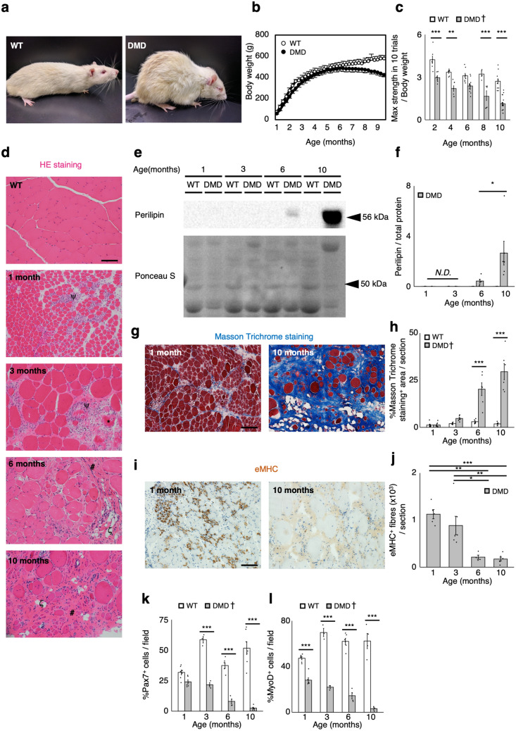
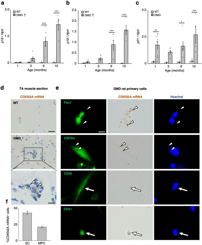
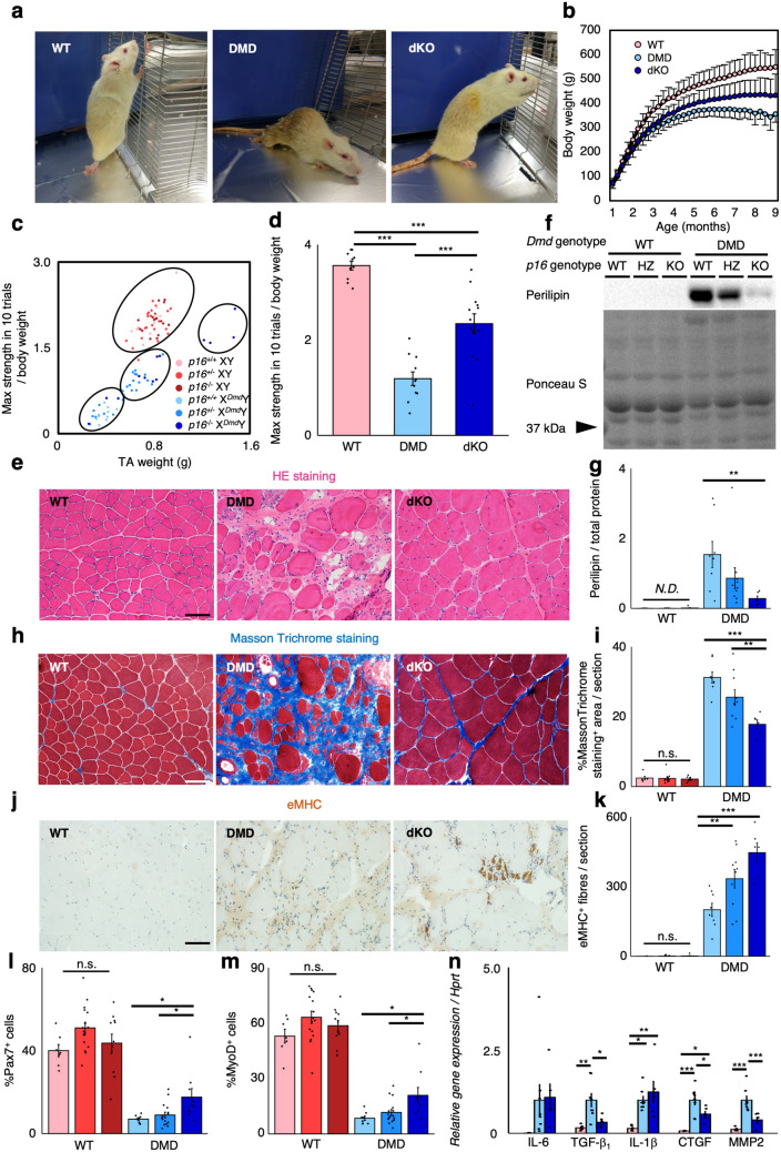
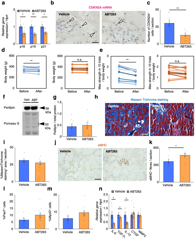
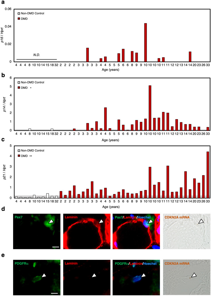
Duchenne muscular dystrophy (DMD) is a progressive disease characterised by chronic muscle degeneration and inflammation. Our previously established DMD model rats (DMD rats) have a more severe disease phenotype than the broadly used mouse model. We aimed to investigate the role of senescence in DMD using DMD rats and patients. Senescence was induced in satellite cells and mesenchymal progenitor cells, owing to the increased expression of CDKN2A, p16- and p19-encoding gene. Genetic ablation of p16 in DMD rats dramatically restored body weight and muscle strength. Histological analysis showed a reduction of fibrotic and adipose tissues invading skeletal muscle, with increased muscle regeneration. Senolytic drug ABT263 prevented loss of body weight and muscle strength, and increased muscle regeneration in rats even at 8 months-the late stage of DMD. Moreover, senescence markers were highly expressed in the skeletal muscle of DMD patients. In situ hybridization of CDKN2A confirmed the expression of it in satellite cells and mesenchymal progenitor cells in patients with DMD. Collectively, these data provide new insights into the integral role of senescence in DMD progression.


Conflict of interest statement

The authors declare no competing interests.

Figures

Figure 1
Figure 1
Progressive exacerbation of muscular dystrophy in DMD rats. (a) Representative images of 6-month-old WT (left panel) and DMD rats (right panel). (b) Body weight comparison of WT and DMD rats of 1–9 months of age (n = 6). (c) Quantification of maximum muscle strength by grip test at the indicated ages of WT and DMD rats (2 months: WT: n = 7, DMD: n = 6, 4 months: WT: n = 7, DMD: n = 7, 6 months: WT: n = 8, DMD: n = 12, 8 months: WT: n = 4, DMD: n = 7, 10 months: WT: n = 11, DMD: n = 21). (d) Representative HE stains of TA muscle sections from 6-month-old WT rats and 1- to 10-month-old DMD rats. Scale bar = 100 μm. The following symbols, ψ, *, #, and ζ, indicate inflammatory cell infiltration, necrotic myofibres, fibrosis, and adipogenesis, respectively. (e) Immunoblotting analysis of perilipin expression in WT and DMD rats. Full-length blots are presented in Supplementary Figure 6a. (f) Quantification of perilipin protein expression (n = 6, each). (g) Representative Masson Trichrome stains of TA muscle sections from 1- to 10-month-old WT and DMD rats. Scale bar = 100 μm. (h) Quantification of Masson Trichrome staining positive area per section (n = 6, each). (i) Immunohistochemical analysis of eMHC in TA muscle sections from WT and DMD rats. Scale bar = 100 μm. (j) Quantification of eMHC positive fibres per section (n = 6, each). (k,l) Quantification of (k) Pax7+ cells and (l) MyoD+ cells of skeletal muscle primary cells from WT and DMD rats (n = 6, each). Data are expressed as mean ± SEM except for (b), which is expressed as mean ± SD, and were compared by Tukey Kramer’s test. Different letters indicate statistically significant differences (p < 0.05). Progressive decrease of both Pax7+ and MyoD+ cells was observed in DMD rats. Significant decrease of the number of MyoD+ cells was observed in DMD rats from 1 month compared to WT, while from 3 months about the number of Pax7+ cells. For (c), (h), (k) and (l), the result of statistical comparison only between the genotypes at each indicated ages was displayed. When a significant age-related difference was observed by the Tukey–Kramer’s test, the † mark was added beside the legend of the graph. *p < 0.05. **p < 0.01. ***p < 0.001. N.D. not detected.
Figure 2
Figure 2
Senescent satellite cells and mesenchymal progenitor cells were present in DMD rats. (ac) Quantification of mRNA levels of senescence markers (p16, p19, and p21) in WT and DMD rat TA muscles (n = 6, each). (d) In situ hybridisation of CDKN2A mRNA using RNAscope on TA muscle sections from 6-month-old WT and DMD rats. Shown are representative images of tissues from WT (top) and DMD rats (middle) with CDKN2A mRNA appearing as brown dots. Scale bar = 50 μm. A higher magnification image from the dotted area is shown in the bottom frame. Scale bar = 10 μm. The white arrowheads show the mononucleated CDKN2A mRNA+ cells. (e) Skeletal muscle primary cells from 6-month-old DMD rats were subjected to CDKN2A mRNA in situ hybridisation using RNAscope before the immunocytochemistry of Pax7, CSPG4, CD45, and CD31. Scale bar = 10 μm. White arrowheads indicate DAB signal. White arrows indicate CD45+ or CD31+ cells. (f) Quantification of CDKN2A+ Pax7+ cells per all Pax7+ cells (shown as SC) and CDKN2A+ CSPG4+ cells per all CSPG4+ cells (shown as MPC) in skeletal muscle primary cells from DMD rats (n = 4, each). Data are expressed as mean + SEM, and were compared by Tukey Kramer’s test. For (ac), the result of statistical comparison only between the genotypes at each indicated ages was displayed. When a significant age-related difference was observed by the Tukey–Kramer’s test, the † mark was added beside the legend of the graph. *p < 0.05. **p < 0.01. ***p < 0.001.
Figure 3
Figure 3
p16KO ameliorates muscular dystrophy in DMD rats. (a) Representative images of 9-month-old WT, DMD, and dKO rats. (b) Body weight comparison of WT, DMD, and dKO rats of 1–9 months of age (n = 20, 23, 20). (c) The plot of maximum muscle strength and TA muscle weight from 9-month-old p16+/+, p16+/−, and p16−/− background WT and DMD rats (n = 10, 22, 17, 11, 24, 13). Clustering was performed using r mclust package. (d) Quantification of maximum muscle strength by grip test in WT, DMD, and dKO rats (n = 10, 11, 13). (e) Representative images of HE stains of TA muscles from 9-month-old WT, DMD, and dKO rats. (f) Immunoblotting analysis of perilipin expression in 9-month-old p16+/+, p16+/−, and p16−/− background WT and DMD rats. Full-length blots are presented in Supplementary Figure 6a. (g) Quantification of perilipin protein expression (n = 6, 13, 8, 8, 10, 6). (h) Masson Trichrome stains of TA muscles from 9-month-old WT, DMD, and dKO rats. (i) Quantification of Masson Trichrome staining positive area per section in 9-month-old p16+/+, p16+/−, and p16−/− background WT and DMD rats (n = 6, 13, 8, 8, 10, 6). (j) Immunohistochemical analysis of eMHC in TA muscle sections from 9-month-old WT, DMD, and dKO rats. (k) Quantification of eMHC positive fibres per section in 9-month-old p16+/+, p16+/−, and p16−/− background WT and DMD rats (n = 6, 13, 8, 8, 10, 6). (l,m) Quantification of (l) Pax7+ and (m) MyoD+ cells of skeletal muscle primary cells from 9-month-old p16+/+, p16+/−, and p16−/− background WT and DMD rats (n = 6, 13, 8, 8, 10, 6). (n) Quantification of mRNA levels of SASP markers (IL-6, TGF-β1, IL-1β, CTGF, and MMP2) in WT, DMD, and dKO rats (n = 6, 8, 6). Data are expressed as mean ± SEM except for (b), which is expressed as mean ± SD, and were compared by Tukey Kramer’s test. For (g), (i), (km), Tukey Kramer’s test was performed only between WT background groups, or between DMD background groups. For (g), (i), (kn), the colour of each bar indicates the genotype as shown in (c). *p < 0.05. **p < 0.01. ***p < 0.001. n.s. not significant; N.D. not detected; scale bar = 100 μm.
Figure 4
Figure 4
Senolytic drug ABT263 inhibits the exacerbation of muscular dystrophy in DMD rats. (a) Quantification of mRNA levels of senescence markers (p16, p19, and p21) in vehicle- and ABT263-treated rats (n = 7, 9). (b) In situ hybridisation of CDKN2A mRNA using RNAscope on TA muscle sections from vehicle- or ABT263-treated rats, with CDKN2A mRNA appearing as red dots. The white arrowheads show the CDKN2A mRNA+ cells. Scale bar = 50 μm. (c) Quantification of the number of CDKN2A mRNA positive mononucleated cells per section from vehicle- or ABT263-treated rats. (d) Body weight comparison before and after treatment with vehicle (left panel) or ABT263 (right panel) (n = 7, 9). (e) Quantification of maximum muscle strength by grip test before and after treatment with vehicle (left panel) or ABT263 (right panel) (n = 7, 9). (f) Immunoblotting analysis of perilipin expression in vehicle- and ABT263-treated rats. Full-length blots are presented in Supplementary Figure 6a. (g) Quantification of perilipin protein expression (n = 7, 9). (h) Masson Trichrome stains of TA muscles from vehicle and ABT263-treated rats. Scale bar = 100 μm. (i) Quantification of Masson Trichrome staining positive area per section (n = 7, 9). (j) Immunohistochemical analysis of eMHC in TA muscle sections from vehicle- and ABT263-treated rats. Scale bar = 100 μm. (k) Quantification of eMHC positive fibres per section (n = 7, 9). (l,m) Quantification of (l) Pax7+ cells and (m) MyoD+ cells of skeletal muscle primary cells from vehicle- and ABT263-treated rats (n = 7, 9). (n) Quantification of mRNA levels of SASP markers (IL-6, TGF-β1, IL-1β, CTGF, and MMP2) in vehicle- and ABT263-treated rats (n = 7, 9). Data are expressed as mean ± SEM. The p-value was determined by paired Student’s t test for (d) and (e), and unpaired Student’s t test for others. *p < 0.05, **p < 0.01. n.s. not significant.
Figure 5
Figure 5
Senescence markers were elevated in human DMD patients. (ac) Quantification of mRNA levels of senescence markers (p16, p14, and p21) in non-DMD control individuals and patients with DMD. Individual data from non-DMD control individuals and DMD patients are expressed as bar graphs (n = 10, 35). The grey band behind the graph indicates the mean ± SEM value range of the non-DMD control group. The figure without a grey band indicate that the target gene expression was not observed in the non-DMD control group. (d,e) Skeletal muscle sections from DMD patients were subjected to in situ hybridisation of CDKN2A mRNA using RNAscope before immunocytochemical analysis of (d) Pax7 or (e) PDGFRα with laminin. CDKN2A mRNA appears as brown dots. Scale bar = 10 μm. White arrowheads indicate DAB signal. N.D. not detected.

References

    1. Ryder S, et al. The burden, epidemiology, costs and treatment for Duchenne muscular dystrophy: an evidence review. Orphanet J. Rare Dis. 2017;12:79. doi: 10.1186/s13023-017-0631-3. - DOI - PMC - PubMed
    1. Mercuri E, Muntoni F. Muscular dystrophies. Lancet. 2013;381:845–860. doi: 10.1016/S0140-6736(12)61897-2. - DOI - PubMed
    1. Yin H, Price F, Rudnicki MA. Satellite cells and the muscle stem cell niche. Physiol. Rev. 2013;93:23–67. doi: 10.1152/physrev.00043.2011. - DOI - PMC - PubMed
    1. Verhaart IEC, Aartsma-Rus A. Therapeutic developments for Duchenne muscular dystrophy. Nat. Rev. Neurol. 2019;15:373–386. doi: 10.1038/s41582-019-0203-3. - DOI - PubMed
    1. Visser M, et al. Relationship of interleukin-6 and tumor necrosis factor-α with muscle mass and muscle strength in elderly men and women: the Health ABC Study. J. Gerontol. Ser. A Biol. Sci. Med. Sci. 2002;57:M326–M332. doi: 10.1093/gerona/57.5.M326. - DOI - PubMed

Publication types

MeSH terms