Review
. 2021 Mar;18(3):169-193.
doi: 10.1038/s41569-020-00435-x. Epub 2020 Oct 12.

Myocarditis and inflammatory cardiomyopathy: current evidence and future directions

Affiliations
Review

Myocarditis and inflammatory cardiomyopathy: current evidence and future directions

Carsten Tschöpe et al. Nat Rev Cardiol. 2021 Mar.

Abstract

Inflammatory cardiomyopathy, characterized by inflammatory cell infiltration into the myocardium and a high risk of deteriorating cardiac function, has a heterogeneous aetiology. Inflammatory cardiomyopathy is predominantly mediated by viral infection, but can also be induced by bacterial, protozoal or fungal infections as well as a wide variety of toxic substances and drugs and systemic immune-mediated diseases. Despite extensive research, inflammatory cardiomyopathy complicated by left ventricular dysfunction, heart failure or arrhythmia is associated with a poor prognosis. At present, the reason why some patients recover without residual myocardial injury whereas others develop dilated cardiomyopathy is unclear. The relative roles of the pathogen, host genomics and environmental factors in disease progression and healing are still under discussion, including which viruses are active inducers and which are only bystanders. As a consequence, treatment strategies are not well established. In this Review, we summarize and evaluate the available evidence on the pathogenesis, diagnosis and treatment of myocarditis and inflammatory cardiomyopathy, with a special focus on virus-induced and virus-associated myocarditis. Furthermore, we identify knowledge gaps, appraise the available experimental models and propose future directions for the field. The current knowledge and open questions regarding the cardiovascular effects associated with severe acute respiratory syndrome coronavirus 2 (SARS-CoV-2) infection are also discussed. This Review is the result of scientific cooperation of members of the Heart Failure Association of the ESC, the Heart Failure Society of America and the Japanese Heart Failure Society.


Conflict of interest statement

C.T. is a consultant for Cardiotropic Labs, Miami, FL, USA. S.B.F. reports grants from Fresenius Medical Care and ENDI Foundation. J.M.H. holds equity in Heart Genomics. J.M.H. and B.H. are both inventors on a patent involving the use of RNA as a biomarker for myocarditis. The other authors declare no competing interests.

Figures

Fig. 1
Fig. 1. Prominent viruses associated with inflammatory cardiomyopathy over time.
Over the years, the number of recognized viruses associated with inflammatory cardiomyopathy has grown. This evolution is partly influenced by the intentional detection of a broader repertoire of viruses over time as well as by the occurrence of novel viruses or virus genotypes in the heart. The association between severe acute respiratory syndrome coronavirus (SARS-CoV) and SARS-CoV-2 and inflammatory cardiomyopathy is not yet clear. ‘(?)’ denotes unclear, needing further investigation; HIV, human immunodeficiency virus. Based on data from ref..
Fig. 2
Fig. 2. Cardiosplenic axis in coxsackievirus B3-induced myocarditis.
In the heart, coxsackievirus B3 infection of cardiomyocytes leads to cell damage and death and the release of IL-1β and damage-associated molecular patterns (DAMPs), which trigger the recruitment and activation of cells from the innate immune system. Pain, anxiety and the release of danger signals into the systemic circulation trigger emergency haematopoiesis in the bone marrow, leading to medullary monocytopoiesis as well as release of myeloid progenitor cells into the circulation. Myeloid progenitor cells then migrate to the spleen, where extramedullary monocytopoiesis takes place to replenish the pool of pro-inflammatory Ly6Chigh monocytes, which can be rapidly mobilized to the damaged heart. In the heart, IFNγ released by infected cardiomyocytes boosts the production by fibroblasts of the pro-inflammatory C-C motif chemokines CCL2 and CCL7, which promote the homing of Ly6Chigh monocytes to the heart. Given that the spleen is a target organ of coxsackievirus B3 and monocytes target cells of coxsackievirus B3, the recruited Ly6Chigh monocytes might be infected with coxsackievirus B3 and thereby transport the virus into the heart, further contributing to the viral infection. Activation of the innate immune system in the heart is beneficial for its antiviral effects but excessive or persistent activation can lead to exaggerated and/or chronic inflammation that triggers myocardial destruction and remodelling, culminating in cardiac dysfunction.
Fig. 3
Fig. 3. Diagnosis of lymphocytic myocarditis.
Acute and healing lymphocytic myocarditis is diagnosed with histology and immunohistology of endomyocardial biopsy samples. a | Acute lymphocytic myocarditis caused by enterovirus A71 infection. Histology image showing cardiomyocyte necrosis (as revealed by the haematoxylin and eosin (H&E) staining in the left panel)) and immunohistology image showing diffuse infiltration of CD3+ T cells (as shown by anti-CD3 antibody staining (brown) in the right panel). b | Healing lymphocytic immune-mediated myocarditis. Histology image showing fibrosis but no cardiomyocyte necrosis (left panel) and immunohistology image showing the presence of infiltrated CD3+ T cells (right panel). All images ×400.
Fig. 4
Fig. 4. Visualization of viral nucleic acids in acute myocarditis.
Viral nucleic acids in heart tissue samples from patients with acute myocarditis can be detected with radioactive in situ hybridization (black spots). Cell nuclei (purple) and cell cytoplasm and extracellular matrix (pink) are visualized with haematoxylin and eosin staining. Enteroviruses (panel a) infect and lyse cardiomyocytes, parvovirus B19 (panel b) infects endothelial cells, and human herpesviruses (panel c) and Epstein–Barr viruses (panel d) replicate in immune cells. Panels a and b ×400, panels c and d ×630.
Fig. 5
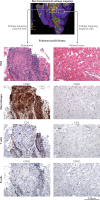
Fig. 5. Electroanatomical voltage mapping to guide endomyocardial biopsy.
Identification of a myocardial scar area with the use of 3D electroanatomical voltage mapping in a patient with suspected cardiac sarcoidosis. The left square in the top panel marks a scar area identified by the presence of low voltages (red and yellow). Subsequent analysis of endomyocardial biopsy samples from this region identified the presence of a sarcoid granuloma in the scar area, visualized with haematoxylin and eosin staining (H&E), with the presence of CD68+ cells (macrophages), CD3+ cells (T cells) and CD20+ cells (B cells), as revealed by antibody staining (brown). By contrast, analysis of endomyocardial biopsy samples from a non-scar area, identified by high voltages (purple) in electroanatomical voltage mapping, showed normal tissue structure and no infiltration of immune cells. All histology images ×100.
Fig. 6
Fig. 6. Improving the subclassification of patients with inflammatory cardiomyopathy.
Clinical characterization of the patient (using risk factors and demographic and quality of life parameters, in addition to basic, physical and laboratory tests, and electrocardiography (ECG), echocardiography and cardiac MRI measurements) — known as phenomapping — combined with histology, immunohistology and viral diagnosis of endomyocardial biopsy samples will allow the classification of patients with inflammatory cardiomyopathy into different clusters, with the ultimate goal of defining therapeutically homogeneous patient subpopulations to improve outcomes. The figure shows a schematic example of a heatmap with hierarchical clustering of the patients on the basis of clinical parameters and endomyocardial biopsy results. GFR, glomerular filtration rate; HIV, human immunodeficiency virus; hsCRP, high-sensitivity C-reactive protein; PvO2, mixed venous oxygen tension.
Fig. 7
Fig. 7. Gaps in evidence for endomyocardial biopsy-guided therapy in myocarditis and inflammatory cardiomyopathy.
Patients with myocarditis or inflammatory cardiomyopathy can be classified into four groups on the basis of endomyocardial biopsy (EMB) results: inflammation-negative, virus-negative; inflammation-positive, virus-negative; inflammation-negative, virus-positive; and inflammation-positive, virus-positive. In patients with virus-positive inflammatory cardiomyopathy, a clear distinction between virus-active and virus-associated inflammatory cardiomyopathy is required. Given the different aetiologies and clinical presentations of the four groups, specific therapy regimens are suggested for each group (blue boxes), in addition to approved optimal medical therapy for heart failure and risk-adjusted therapy. Immunosuppressive therapy is mandatory for specific forms of virus-negative autoimmune myocarditis, such as eosinophilic myocarditis, giant-cell myocarditis and cardiac sarcoidosis. Immunosuppressive therapy is also safe and effective in clinically unstable or non-resolving lymphocytic virus-negative myocarditis and in lymphocytic virus-negative myocarditis refractory to standard heart failure therapy. Autoantibody targeting can be achieved with immunoadsorption or with newly developed small molecules (aptamers) that neutralize specific autoantibodies. Autoantibody targeting is also under investigation for the treatment of non-primary inflammatory heart diseases, in which autoimmunity could have a role in disease progression. However, knowledge gaps remain about the type and length of immunosuppression and on novel biological agents to target specific immune pathways or autoantibodies. Data from registries and large randomized clinical trials are needed to evaluate the efficacy of the different proposed regimens, which will contribute to improving the clinical value of EMB-guided diagnosis. The role of severe acute respiratory syndrome coronavirus 2 (SARS-CoV-2) infection in myocarditis and corresponding treatment options are still unclear; therefore, SARS-CoV-2 is not included in the figure. (?) denotes unclear, needs further investigation; B19V, parvovirus B19; HCV, hepatitis C virus; IVIG, intravenous immunoglobulins.

References

    1. Richardson P, et al. Report of the 1995 World Health Organization/International Society and Federation of Cardiology task force on the definition and classification of cardiomyopathies. Circulation. 1996;93:841–842. doi: 10.1161/01.CIR.93.5.841. - DOI - PubMed
    1. Caforio AL, et al. Current state of knowledge on aetiology, diagnosis, management, and therapy of myocarditis: a position statement of the European Society of Cardiology Working Group on Myocardial and Pericardial Diseases. Eur. Heart J. 2013;34:2636–2648. doi: 10.1093/eurheartj/eht210. - DOI - PubMed
    1. Ammirati E, et al. Clinical presentation and outcome in a contemporary cohort of patients with acute myocarditis. Circulation. 2018;138:1088–1099. doi: 10.1161/CIRCULATIONAHA.118.035319. - DOI - PubMed
    1. Kociol RD, et al. Recognition and initial management of fulminant myocarditis: a scientific statement from the American Heart Association. Circulation. 2020;141:e69–e92. doi: 10.1161/CIR.0000000000000745. - DOI - PubMed
    1. Ammirati E, et al. Fulminant versus acute nonfulminant myocarditis in patients with left ventricular systolic dysfunction. J. Am. Coll. Cardiol. 2019;74:299–311. doi: 10.1016/j.jacc.2019.04.063. - DOI - PubMed

MeSH terms