. 2020 Dec;27(12):1142-1151.
doi: 10.1038/s41594-020-0510-9. Epub 2020 Oct 12.

The structure of a calsequestrin filament reveals mechanisms of familial arrhythmia

Affiliations

The structure of a calsequestrin filament reveals mechanisms of familial arrhythmia

Erron W Titus et al. Nat Struct Mol Biol. 2020 Dec.

Abstract

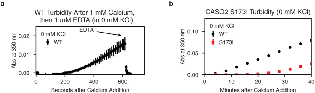
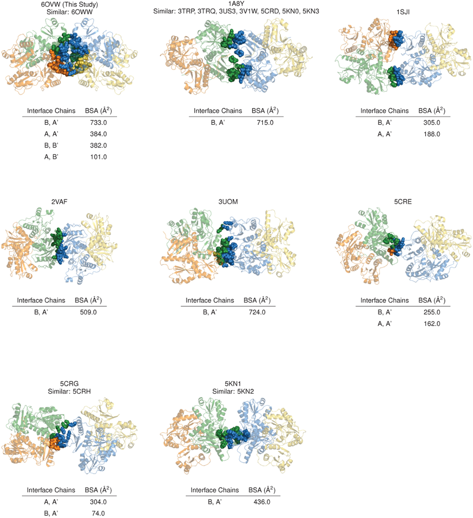
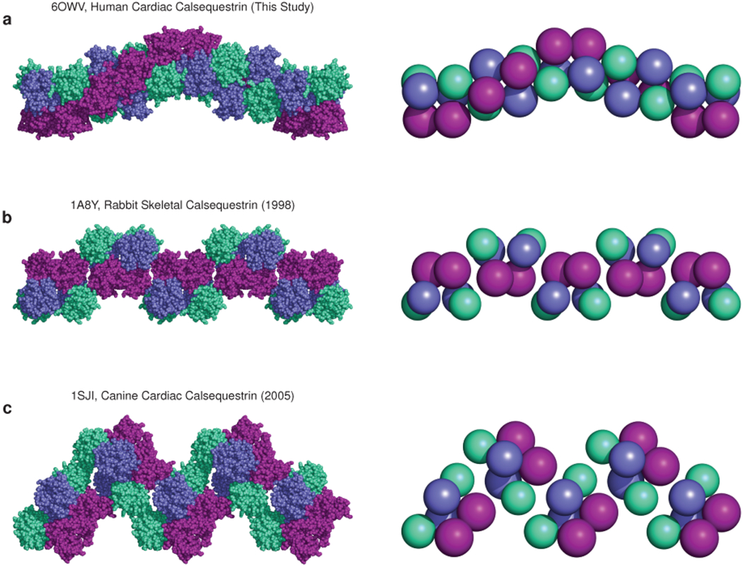
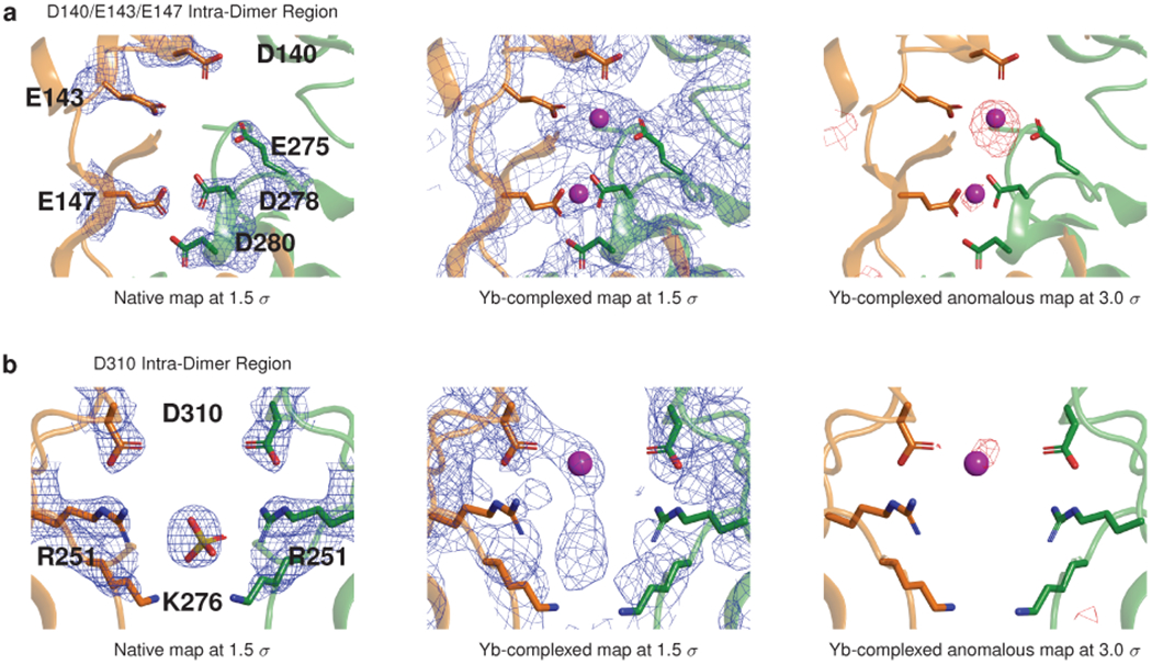
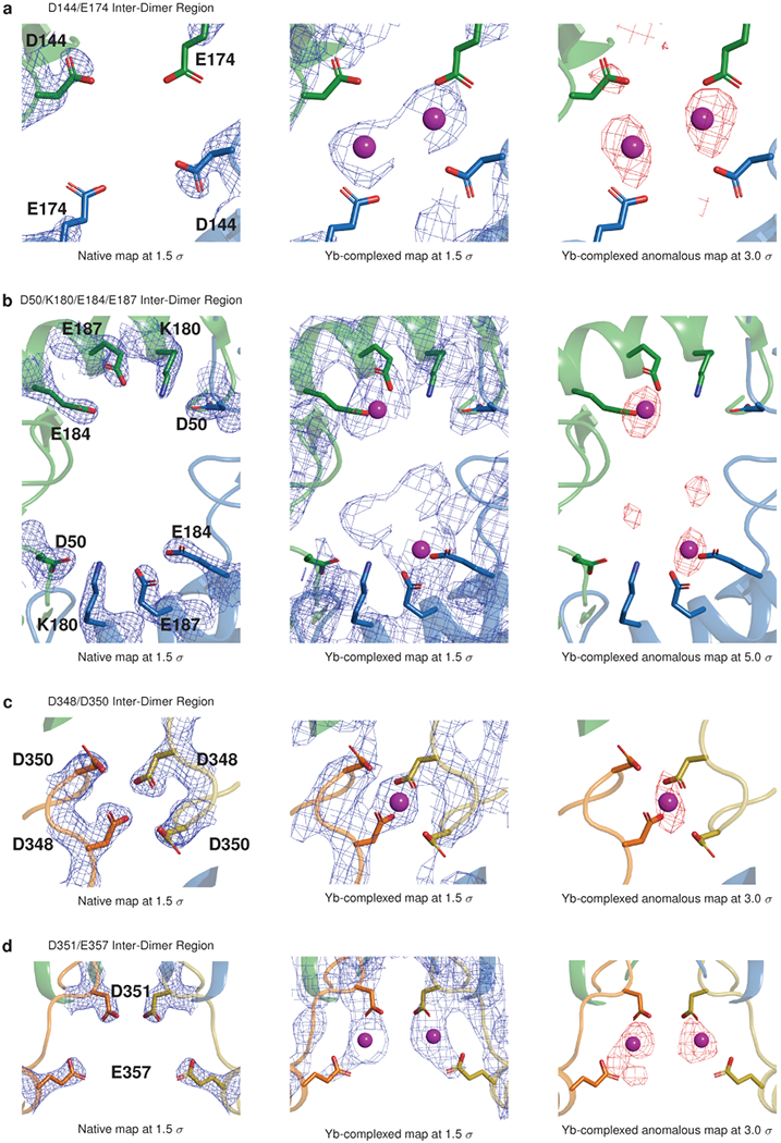
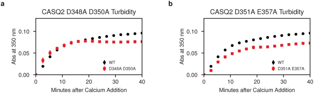
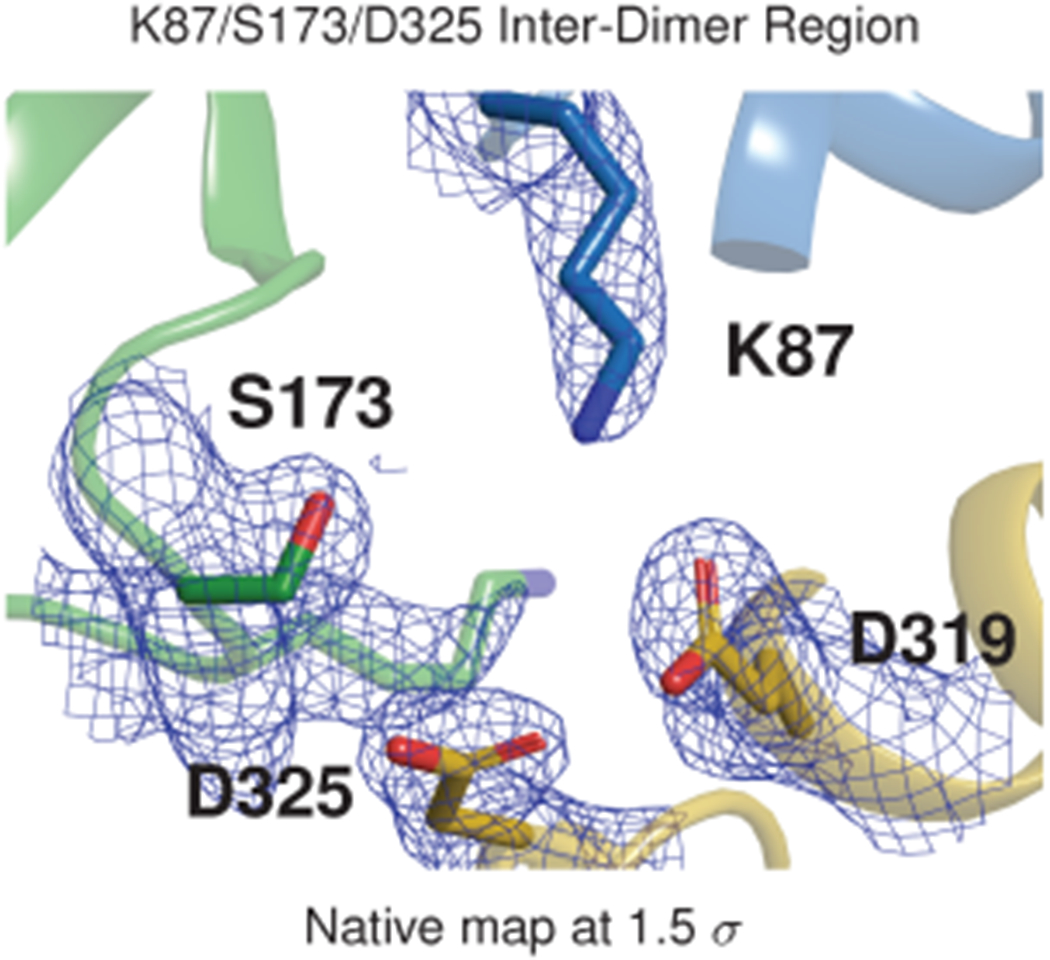
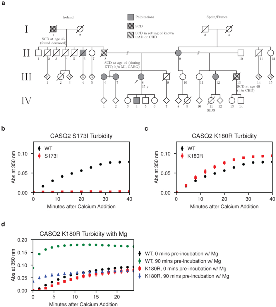
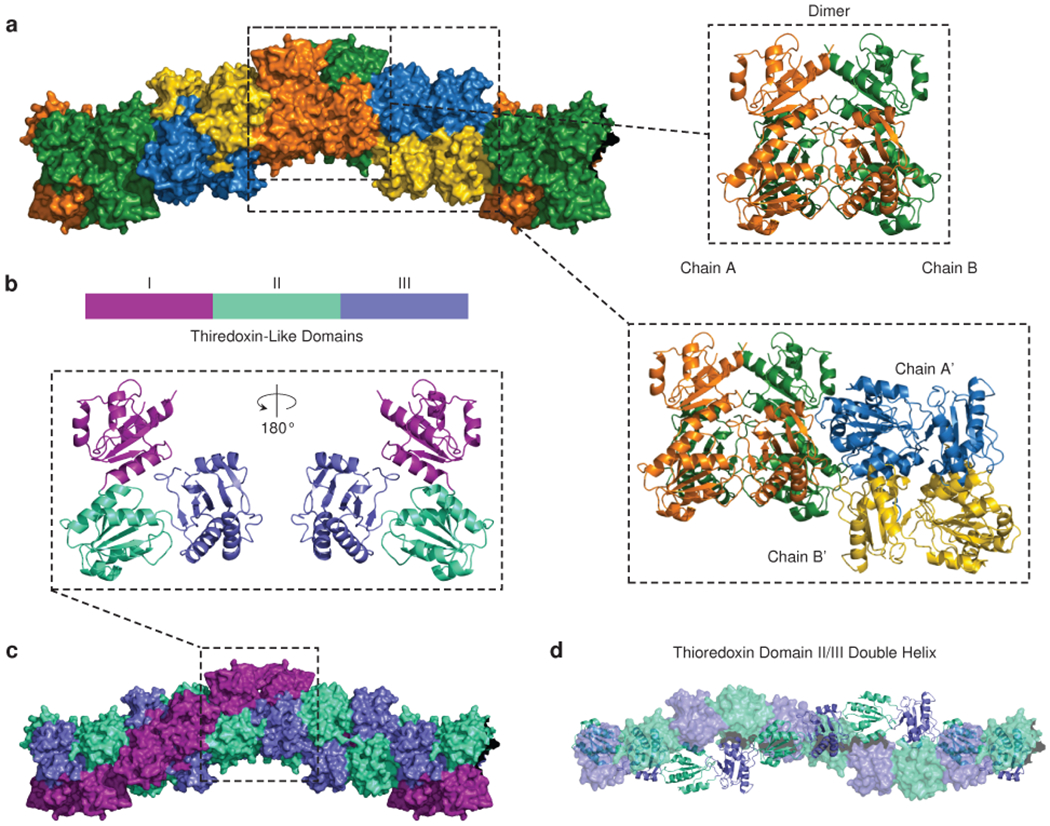
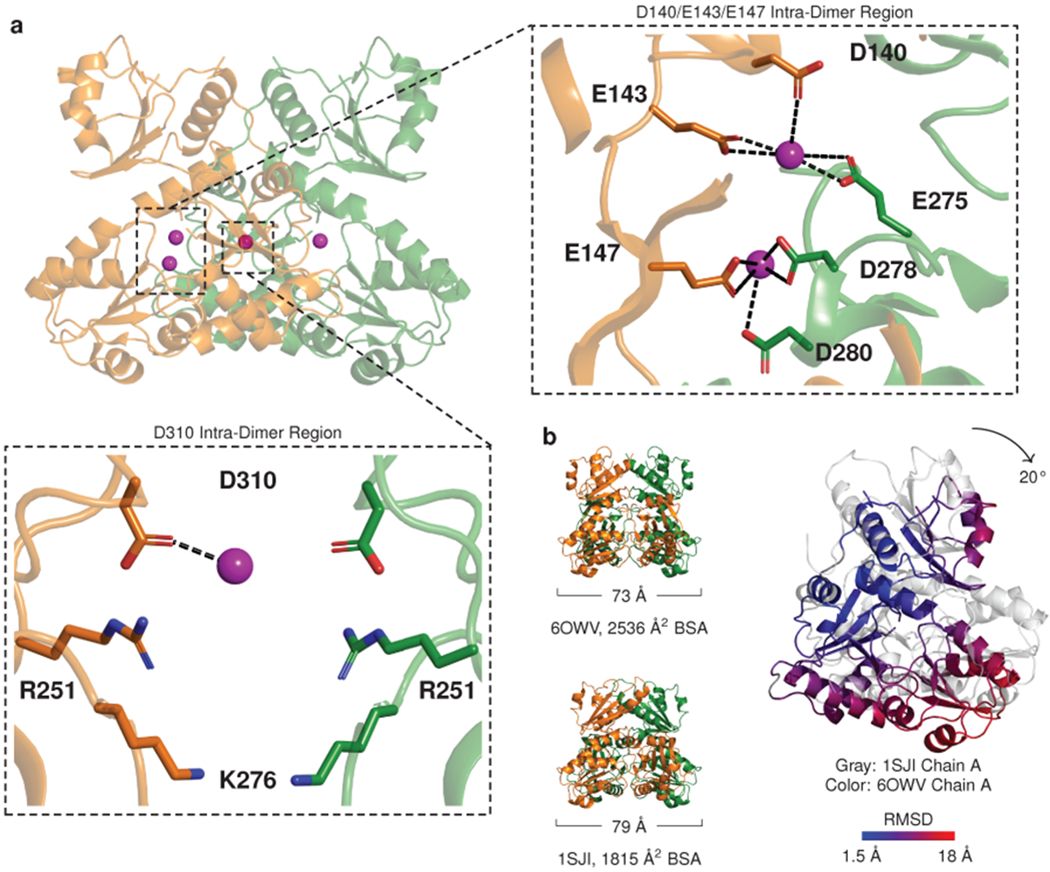
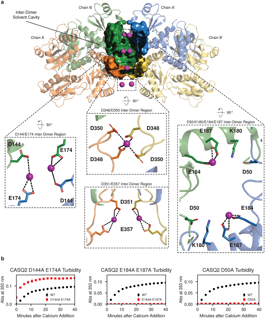
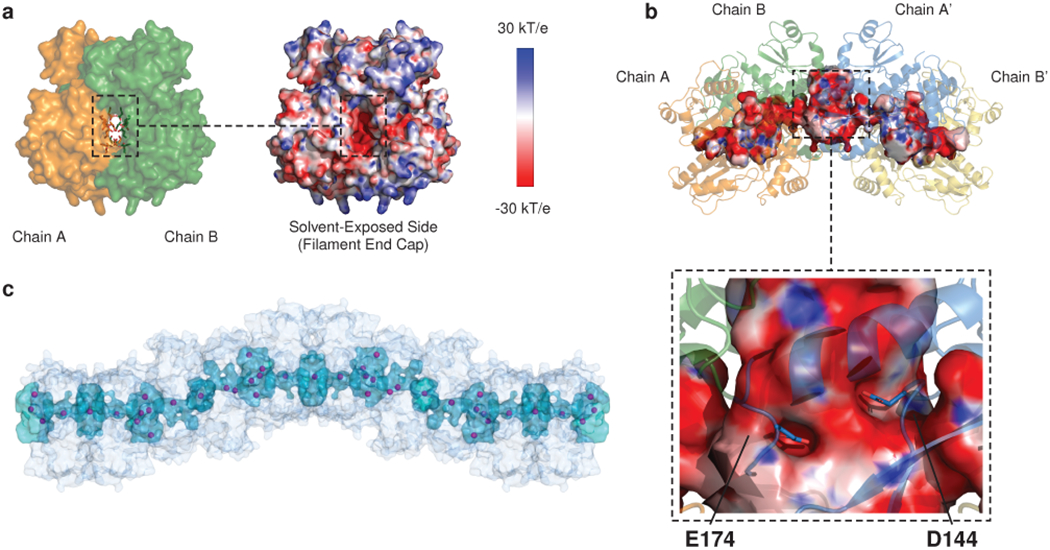
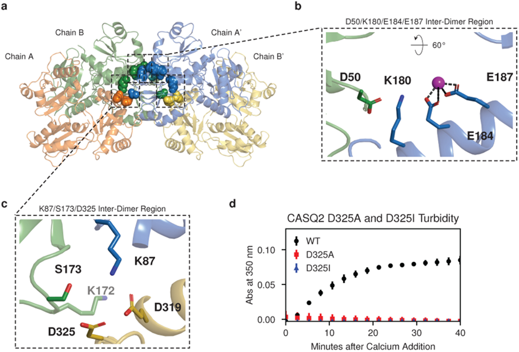
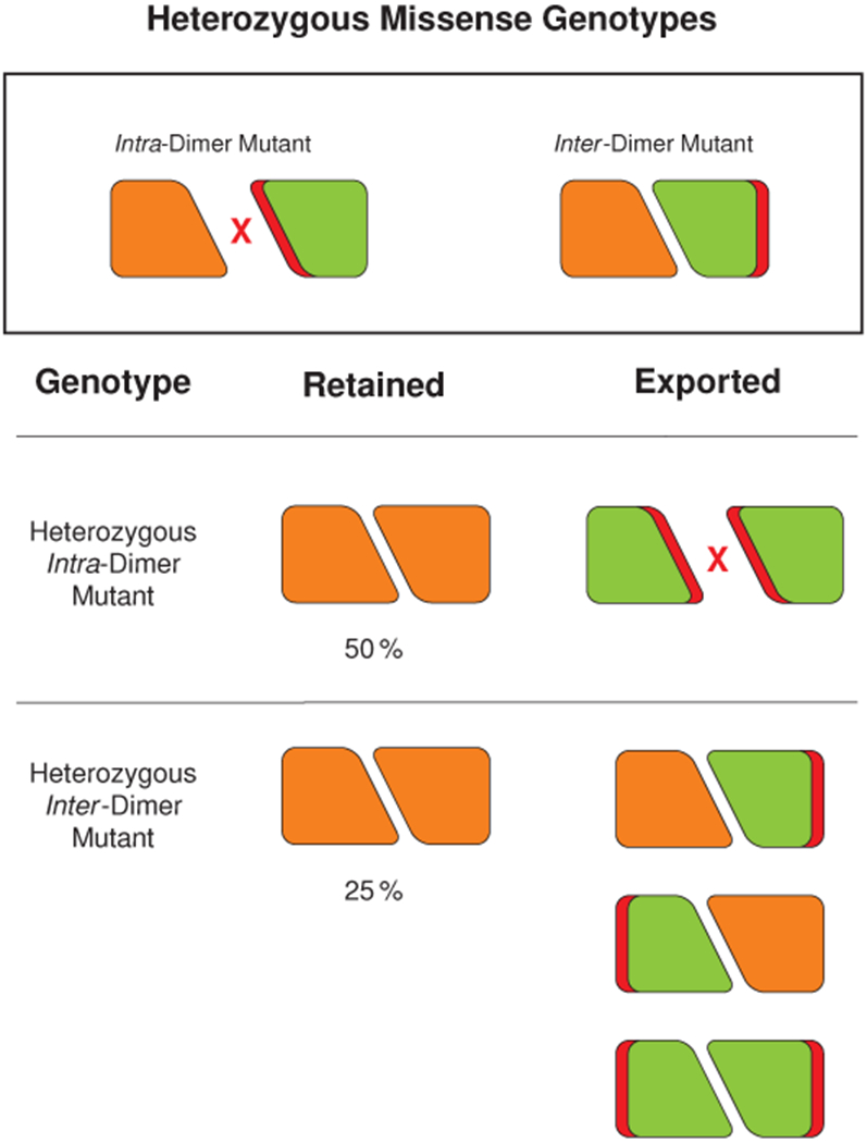
Mutations in the calcium-binding protein calsequestrin cause the highly lethal familial arrhythmia catecholaminergic polymorphic ventricular tachycardia (CPVT). In vivo, calsequestrin multimerizes into filaments, but there is not yet an atomic-resolution structure of a calsequestrin filament. We report a crystal structure of a human cardiac calsequestrin filament with supporting mutational analysis and in vitro filamentation assays. We identify and characterize a new disease-associated calsequestrin mutation, S173I, that is located at the filament-forming interface, and further show that a previously reported dominant disease mutation, K180R, maps to the same surface. Both mutations disrupt filamentation, suggesting that disease pathology is due to defects in multimer formation. An ytterbium-derivatized structure pinpoints multiple credible calcium sites at filament-forming interfaces, explaining the atomic basis of calsequestrin filamentation in the presence of calcium. Our study thus provides a unifying molecular mechanism through which dominant-acting calsequestrin mutations provoke lethal arrhythmias.


Conflict of interest statement

Declaration of Interests

The authors declare no competing interests.

Figures

Extended Data Figure 1:
Extended Data Figure 1:. Turbidity assay controls; related to Fig. 1.
a, Stoichiometric addition of EDTA demonstrates immediate reversal of calcium-induced turbidity. b, Turbidity assay for the S173I mutant in 0 mM KCl. Error bars represent the mean ± s.d of n=3 technical replicates.
Extended Data Figure 2:
Extended Data Figure 2:. The inter-dimer interface of the new candidate cardiac calsequestrin filament exhibits all-by-all contacts and greater buried surface area (BSA) compared to all other published calsequestrin structures; related to Fig. 2.
For each published calsequestrin structure, the inter-dimer interface with the greatest buried surface area is shown. Residues with buried surface area at the interface are rendered as spheres. Where similar PDB codes are listed, inter-dimer interfaces are roughly isomorphous with the example structure shown, although the space group and unit cell used to determine the structure sometimes differ.
Extended Data Figure 3:
Extended Data Figure 3:. The 3-Helix configuration of the new filament candidate promotes close packing of thioredoxin domains.
a, The candidate cardiac calsequestrin filament assembled from crystallographic symmetry operations on PDB ID 6OWV (human CASQ2, this study). The filament exhibits tight packing of protomers and thioredoxin domains (shown on the right using equal-size spheres placed at the center of mass of each thioredoxin domain). b, A putative skeletal calsequestrin filament assembled from crystallographic symmetry operations on PDB ID 1A8Y (rabbit CASQ1, 1998). Right-side: equal-size spheres represent thioredoxin domains. c, A putative skeletal calsequestrin filament assembled from crystallographic symmetry operations on PDB ID 1SJI (canine CASQ2, 2005). Right-side: equal-size spheres represent thioredoxin domains.
Extended Data Figure 4:
Extended Data Figure 4:. Electron density and anomalous difference maps for Yb-binding sites at the cardiac calsequestrin intra-dimer interface; related to Fig. 3.
a, Electron density (blue mesh) and anomalous difference maps for the D140-E143-E147 region. b, Electron density and anomalous difference maps for the D310 region.
Extended Data Figure 5:
Extended Data Figure 5:. Dimer overlays reveal that calsequestrin structures can be classified into tightly packed or loosely packed dimers; related to Fig. 3.
Dimers from published calsequestrin structures (lighter orange and green) are overlaid onto the tightly packed dimer from the present study (6OVW, darker orange and green). In each dimer pair, chain A is aligned to chain A to illustrate the relative displacement of chain B. The concentration of divalent cations used in the crystallization conditions is noted below. The overlays reveal two distinct conformational groupings. The more tightly packed conformation with inwardly rotated chains resembles the dimer in this study and appears to form at low pH or in the presence of neutralizing divalents.
Extended Data Figure 6:
Extended Data Figure 6:. Tightly packed calsequestrin dimers consistently exhibit increased conformational disorder in domain I; related to Fig. 3.
The top panel shows tightly packed calsequestrin dimers (i.e. dimers of calsequestrin crystallized in low pH or with high concentration of multivalent cations). In these structures, solvent-exposed loops in domain I are consistently disordered. In PDB 6OVW, the disordered loop region is omitted due to the high level of disorder. This same region (boxed, residues 58-68) is highly disordered in similar structures. The bottom panel shows loosely packed calsequestrin dimers (i.e. dimers of calsequestrin crystallized at neutral pH with low or trace concentrations of multivalent cations). The resolution for each structure is indicated, and several structures of non-comparable resolution are excluded (2VAF, 5CRE, 5KN0).
Extended Data Figure 7:
Extended Data Figure 7:. Electron density and anomalous difference maps for Yb-binding Sites at the cardiac calsequestrin filament’s inter-dimer interface; related to Fig. 4.
a, Electron density and anomalous difference maps for the D144-E174 region of interest. b, Electron density and anomalous difference maps for the D50-K180-E184-E187 region. c, Electron density and anomalous difference maps for the D348-D350 region. d, Electron density and anomalous difference maps for the D351-E357 region.
Extended Data Figure 8:
Extended Data Figure 8:. Turbidity assays showing effect of alanine mutagenesis of additional Yb-binding sites at the cardiac calsequestrin inter-dimer interface; related to Fig. 4.
a, Turbidity assay after alanine mutagenesis of the putative calcium-coordinating residues D348 and D350. b, Turbidity assay after alanine mutagenesis of the putative calcium-coordinating residues D351 and E357. Error bars represent the mean ± s.d of n=3 technical replicates.
Extended Data Figure 9:
Extended Data Figure 9:. Electron density map for a hydrophilic pocket at the cardiac calsequestrin filament’s inter-dimer interface; related to Fig. 6.
The S173 inter-dimer region of the calsequestrin filament with electron density shown as a blue mesh.
Fig. 1:
Fig. 1:. Autosomal dominant CASQ2 disease mutations disrupt calsequestrin multimerization.
a, Pedigree of a large extended family with the S173I mutation and a CPVT-like phenotype. CABG = coronary artery bypass graft; CAD = coronary artery disease; CHD = congenital heart disease; ETT = exercise treadmill test; MI = myocardial infarction; SCD = sudden cardiac death; SIDS = sudden infant death syndrome. b, Multimerization kinetics of purified S173I mutant protein assessed by turbidity assays following addition of 1 mM CaCl2 (see Methods for details). c, Multimerization kinetics of the K180R mutant determined under the same conditions as in (b). d, Multimerization kinetics of the K180R mutant observed using a turbidity assay (same conditions as in b, but with 2 mM MgCl2 added prior to calcium). Error bars represent the mean ± s.d. of 3 technical replicates.
Fig. 2:
Fig. 2:. Helical domain architecture of the cardiac calsequestrin filament.
a, A space-filling model of the cardiac calsequestrin candidate filament (PDB ID 6OWV) is shown, along with ribbon diagrams of a dimeric and a tetrameric assembly (boxed images). Dimers are stacked on a screw axis with 90 degrees of rotation per dimer. b, Cardiac calsequestrin monomers are colored by thioredoxin domain. The monomers are translated but remain in their dimer-forming orientation. c, The helical character of the filament is revealed at the domain level. Viewed at the level of its thioredoxin domains (3 per protomer), the filament consists of an inner thioredoxin double helix (domains II and III) with an outer thioredoxin single helix (domain I) wrapping the double helical core. d, The inner double helix of the filament consists of thioredoxin domain II and III. For clarity, one strand is rendered as ribbon (domains from chains A and A’), while the other strand is rendered as surface (domains from chains B and B’).
Fig. 3:
Fig. 3:. Cation binding leads to conformational shifts in calsequestrin dimers.
a, Ribbon diagram of a dimer with ytterbium (Yb) sites (magenta spheres) within its interior cavity. Boxed zoom images highlight Yb positions that bridge dimer chains A and B: relatively high occupancy is observed in a narrow intra-dimer cleft (right panel), while a site of weaker interaction and weaker occupancy is identified in the intra-dimer cavity (lower panel). b, Comparison of a previously reported cardiac calsequestrin dimer (1SJI) to the more tightly packed dimer of the present study. The tightly packed dimer results primarily from rigid body rotation of the dimer chains inward (for a single chain, a 20° counterclockwise rotation is observed in the plane of the page when the other chain is fixed to the reference dimer). The inward rotation produces an increase in buried surface area (BSA) in thioredoxin domains II and III.
Fig. 4:
Fig. 4:. Cations are trapped at inter-dimer filament-forming interfaces.
a, Yb (magenta spheres) bound within the walled pocket formed by the inter-dimer interface, with boxed zoomed images highlighting ytterbium sites at D144 and E174, E184 and E187, D348 and D350, and D351 and D357. Thioredoxin domain II of chain A’ (blue) is omitted to allow visualization of the interior of the solvent pocket formed by the inter-dimer interface. b, Turbidity assays after alanine mutagenesis of putative calcium-binding and salt bridge residues. Left: D144A and E174A. Middle: E184A and E187A. Right: D50A. Error bars indicate the mean ± s.d. of 3 technical replicates.
Fig. 5:
Fig. 5:. The cardiac calsequestrin filament contains a continuous, solvent-accessible lumen along its long axis.
a, Left: space-filling model of the interior cavity of the dimer viewed down its long axis. Residues that interact with Yb atoms within the intra-dimer cleft (Fig. 3) are shown as sticks. All other residues are rendered as surface. Right: APBS-generated electrostatic surface charge distribution of the same region. b, The lumen is continuous down the length of filament because of the large solvent cavity formed at each dimer-dimer interface. The APBS-generated electrostatic surface of the lumen (traced by HOLLOW using a 1.4 Å probe) is shown, with closeup of residues D144 and E174 deep within the electronegative cavity. c, View of the filament and its continuous interior cavity, with Yb sites shown as magenta spheres.
Fig. 6:
Fig. 6:. Dominant disease-associated mutations disrupt the filament-forming interface.
a, Inter-dimer interface residues in the vicinity of S173 and K180 are shown as spheres colored as in Fig 2a. b, Closeup of K180 with the adjacent E184 and E187 cation binding site, with the coordinated Yb atom shown as a magenta sphere. c, Hydrophilic pocket at S173 created by interactions of 3 different thioredoxin domains from 3 distinct chains (K87, S173, D325). K172 (light gray label) is the most proximal of several residues that shield the pocket from bulk solvent. d, Turbidity assay with the D325A and D325I mutations. Error bars indicate the mean ± s.d. of 3 technical replicates.
Fig. 7:
Fig. 7:. Heterozygous mutations at different calsequestrin interfaces display different disease inheritance patterns.
Mutations that inhibit dimerization are likely to cause penetrant disease only when carried recessively - a consequence of export of mutant monomers from the SR. However, mutations that inhibit inter-dimer interaction are likely to have dominant effects - a consequence of the fact that only 25 % of dimers remain able to participate in filament formation.

Similar articles

Cited by

References

    1. Bers DM Macromolecular complexes regulating cardiac ryanodine receptor function. J. Mol. Cell. Cardiol 37, 417–429 (2004). - PubMed
    1. Royer L & Ríos E Deconstructing calsequestrin. Complex buffering in the calcium store of skeletal muscle. J. Physiol 587, 3101–3111 (2009). - PMC - PubMed
    1. Guerrero-Hernández A et al. in Calcium Signaling (ed Islam MS) 337–370 (Springer International Publishing, 2020).
    1. MacLennan DH & Wong PT Isolation of a calcium-sequestering protein from sarcoplasmic reticulum. Proc. Natl. Acad. Sci. U. S. A 68, 1231–1235 (1971). - PMC - PubMed
    1. MacLennan DH Isolation of a second form of calsequestrin. J. Biol. Chem 249, 980–984 (1974). - PubMed

Methods References

    1. Winter G et al. DIALS: implementation and evaluation of a new integration package. Acta Crystallogr D Struct Biol 74, 85–97 (February 2018). - PMC - PubMed
    1. Winter G xia2: an expert system for macromolecular crystallography data reduction. J. Appl. Crystallogr 43, 186–190 (2010).
    1. Evans PR & Murshudov GN How good are my data and what is the resolution? Acta Crystallogr. D Biol. Crystallogr 69, 1204–1214 (2013). - PMC - PubMed
    1. Evans P Scaling and assessment of data quality. Acta Crystallogr. D Biol. Crystallogr 62, 72–82 (2006). - PubMed
    1. Winn MD et al. Overview of the CCP4 suite and current developments. Acta Crystallogr. D Biol. Crystallogr 67, 235–242 (2011). - PMC - PubMed

Publication types

MeSH terms