Review
. 2020 Nov;53(11):e12915.
doi: 10.1111/cpr.12915. Epub 2020 Oct 13.

Programmed cell death pathways in hearing loss: A review of apoptosis, autophagy and programmed necrosis

Affiliations
Review

Programmed cell death pathways in hearing loss: A review of apoptosis, autophagy and programmed necrosis

Junhao Wu et al. Cell Prolif. 2020 Nov.

Abstract

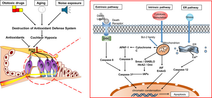
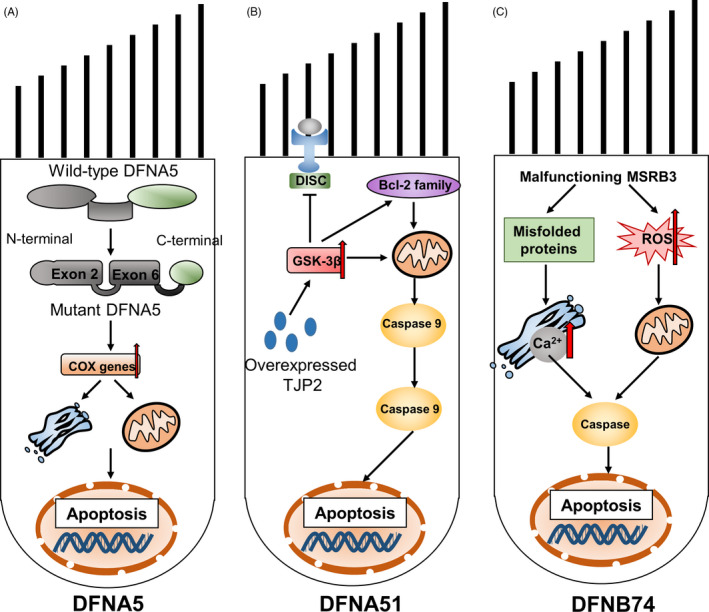
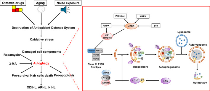
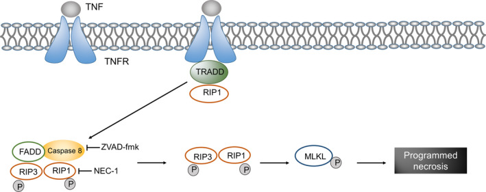
Programmed cell death (PCD)-apoptosis, autophagy and programmed necrosis-is any pathological form of cell death mediated by intracellular processes. Ototoxic drugs, ageing and noise exposure are some common pathogenic factors of sensorineural hearing loss (SNHL) that can induce the programmed death of auditory hair cells through different pathways, and eventually lead to the loss of hair cells. Furthermore, several mutations in apoptotic genes including DFNA5, DFNA51 and DFNB74 have been suggested to be responsible for the new functional classes of monogenic hearing loss (HL). Therefore, in this review, we elucidate the role of these three forms of PCD in different types of HL and discuss their guiding significance for HL treatment. We believe that further studies of PCD pathways are necessary to understand the pathogenesis of HL and guide scientists and clinicians to identify new drug targets for HL treatment.

Keywords: apoptosis; autophagy; hearing loss; programmed necrosis.


Conflict of interest statement

The authors declare no conflict of interest.

Figures

FIGURE 1
FIGURE 1
Apoptotic signalling pathways. Factors such as ototoxic drugs, ageing and noise exposure, which lead to hearing loss, damage the antioxidant defence system of the cochlea and cause imbalance of oxidation‐reduction in the inner ear. Reactive oxygen species (ROS) can directly induce the intrinsic apoptosis of cells. Moreover, they can induce the production of cell death ligands to mediate the extrinsic apoptosis process. ROS‐induced intracellular protein damage can cause endoplasmic reticulum stress, which can lead to apoptosis
FIGURE 2
FIGURE 2
Schematics of three mutations that lead to monogenic hearing loss. A, DFNA5: the apoptosis‐inducing region of DFNA5 is located in exons 2 and 6 of the N‐terminal domain. Skipping exon 8 can change and shorten the C‐terminal domain of DFNA5, reveal the apoptosis‐inducing region and lead to apoptosis. B, DFNA51: overexpression of TJP2 induces apoptosis by activating glycogen synthase kinase 3β (GSK‐3β). C, DFNB74: malfunctioning MSRB3 leads to the accumulation of oxidative damage proteins and reactive oxygen species (ROS), ultimately leading to endoplasmic reticulum stress and subsequent activation of endogenous apoptotic pathways
FIGURE 3
FIGURE 3
Signalling pathway of autophagy in HL. Unc‐51‐like autophagy activating kinase 1 (ULK1) is phosphorylated upon the activation of AMPK and inactivation of mTOR. The activated ULK1 complex and class III phosphoinositide 3 kinase (PI3K) complex form phosphors. Microtubule‐associated protein 1 light‐chain 3 (LC3) protein can be coupled with phosphatidylethanolamine to form LC3Ⅱ. The complex of ATG5, ATG12, ATG16L and LC3Ⅱ can stimulate the elongation of phagocytes, which provide a platform for the formation of phagosomes. On approaching the ubiquitin protein binding to p60 and LC3Ⅱ, the phagosome closes to form autophagosomes. Further, autophagosomes fuse with lysosomes to form autolysosomes in which the contents are degraded
FIGURE 4
FIGURE 4
Signalling pathway of programmed necrosis in hearing loss. When a ligand binds to tumour necrosis factor receptor (TNFR), a combination of TNFR‐associated death domain (TRADD) and receptor‐interacting protein (RIP) 1 increases the level of RIP3 and induces self‐ and transphosphorylation, which is followed by the oligomerization of phosphorylated RIP3. Active RIP3 catalyses the phosphorylation of the mixed lineage kinase domain‐like protein (MLKL), thus resulting in the formation of MLKL oligomers and in translocation to the plasma membrane. Through the reversal mechanism, specific phosphatidylinositol phosphates are combined that lead to plasma membrane permeability and eventually cell necrosis

References

    1. Marazita ML, Ploughman LM, Rawlings B, Remington E, Arnos KS, Nance WE. Genetic epidemiological studies of early‐onset deafness in the U.S. school‐age population. Am J Med Genet Part A. 1993;46(5):486‐491. 10.1002/ajmg.1320460504 - DOI - PubMed
    1. Huth ME, Ricci AJ, Cheng AG. Mechanisms of aminoglycoside ototoxicity and targets of hair cell protection. International J Otolaryngol. 2011;2011:937861 10.1155/2011/937861 - DOI - PMC - PubMed
    1. Chen H, Tang J. The role of mitochondria in age‐related hearing loss. Biogerontology. 2014;15(1):13‐19. 10.1007/s10522-013-9475-y - DOI - PubMed
    1. Hong O, Kerr MJ, Poling GL, Dhar S. Understanding and preventing noise‐induced hearing loss. Disease‐a‐month. 2013;59(4):110‐118. 10.1016/j.disamonth.2013.01.002 - DOI - PubMed
    1. Green DR, Llambi F. Cell death signaling. Cold Spring Harbor Perspect Biol. 2015;7(12). 10.1101/cshperspect.a006080. - DOI - PMC - PubMed