

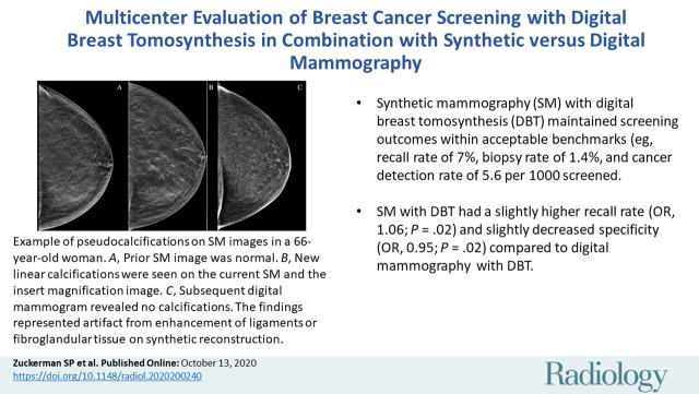
Multicenter Evaluation of Breast Cancer Screening with Digital Breast Tomosynthesis in Combination with Synthetic versus Digital Mammography
- PMID: 33048032
- PMCID: PMC7706877
- DOI: 10.1148/radiol.2020200240
Multicenter Evaluation of Breast Cancer Screening with Digital Breast Tomosynthesis in Combination with Synthetic versus Digital Mammography
Abstract
BackgroundDigital breast tomosynthesis (DBT) combined with digital mammography (DM) is increasingly used in the United States instead of DM alone for breast cancer screening. Early screening outcomes incorporating synthetic mammography (SM) with DBT have suggested that SM is an acceptable non-radiation dose alternative to DM.PurposeTo compare multicenter outcomes from breast cancer screening with SM/DBT versus DM/DBT.Materials and MethodsThis was a retrospective study of consecutive screening mammograms obtained at two institutions. Eligible studies consisted of 34 106 DM/DBT examinations between October 3, 2011, and October 31, 2014, and 34 180 SM/DBT examinations between January 7, 2015, and February 2, 2018, at the University of Pennsylvania and 51 148 DM/DBT examinations between January 1, 2012, and May 31, 2016, and 31 929 SM/DBT examinations between June 1, 2016, and March 30, 2018, at the University of Vermont. Demographics of women who attended screening and results from screening were recorded. Recall rate, biopsy rate, false-negative rate, cancer detection rate, positive predictive value, sensitivity, and specificity were calculated according to modality and institution. Descriptive statistics, χ2 tests, and logistic regression were used in analysis.ResultsThe study included 151 363 screening examinations among 151 363 women (mean age, 58.1 years ± 10.9 [standard deviation]). The unadjusted recall rate was lower with SM/DBT than with DM/DBT (7.0% [4630 of 66 109 examinations] for SM/DBT vs 7.9% [6742 of 85 254 examinations] for DM/DBT; P < .01). However, after multivariable adjustment, SM/DBT was associated with a slightly higher recall rate compared with DM/DBT (adjusted odds ratio [OR], 1.06; adjusted 95% CI: 1.01, 1.11; P = .02). Similarly, after multivariable adjustment, SM/DBT was associated with slightly lower specificity compared with DM/DBT (adjusted OR, 0.95; adjusted 95% CI: 0.90, 0.99; P = .02). There was no statistically significant difference in biopsy rate (P = .54), false-negative rate (P = .38), cancer detection rate (P = .55), invasive or in situ cancer detection rate (P = .52 and P = .98, respectively), positive predictive value (P = .78), or sensitivity (P = .33) for SM/DBT versus DM/DBT overall or within either institution (P > .05 for all).ConclusionBreast cancer screening performance is maintained within benchmarks when synthetic mammography replaces digital mammography in digital breast tomosynthesis imaging.© RSNA, 2020Online supplemental material is available for this article.See also the editorial by Lång in this issue.
Figures
Comment in
-
Mounting Evidence for Synthetic Mammography in Breast Cancer Screening.Radiology. 2020 Dec;297(3):554-555. doi: 10.1148/radiol.2020203716. Epub 2020 Oct 13. Radiology. 2020. PMID: 33064034 No abstract available.
References
-
- Hardesty LA, Kreidler SM, Glueck DH. Digital Breast Tomosynthesis Utilization in the United States: A Survey of Physician Members of the Society of Breast Imaging. J Am Coll Radiol 2016;13(11S):R67–R73. - PubMed
-
- Gao Y, Babb JS, Toth HK, Moy L, Heller SL. Digital Breast Tomosynthesis Practice Patterns Following 2011 FDA Approval: A Survey of Breast Imaging Radiologists. Acad Radiol 2017;24(8):947–953. - PubMed
-
- Friedewald SM, Rafferty EA, Rose SL, et al. Breast cancer screening using tomosynthesis in combination with digital mammography. JAMA 2014;311(24):2499–2507. - PubMed
Publication types
MeSH terms
Grants and funding
LinkOut - more resources
Full Text Sources
Medical
