Review
. 2020 Nov;19(11):e13260.
doi: 10.1111/acel.13260. Epub 2020 Oct 13.

Thyroid hormones in diabetes, cancer, and aging

Affiliations
Review

Thyroid hormones in diabetes, cancer, and aging

Benoit R Gauthier et al. Aging Cell. 2020 Nov.

Abstract

Thyroid function is central in the control of physiological and pathophysiological processes. Studies in animal models and human research have determined that thyroid hormones modulate cellular processes relevant for aging and for the majority of age-related diseases. While several studies have associated mild reductions on thyroid hormone function with exceptional longevity in animals and humans, alterations in thyroid hormones are serious medical conditions associated with unhealthy aging and premature death. Moreover, both hyperthyroidism and hypothyroidism have been associated with the development of certain types of diabetes and cancers, indicating a great complexity of the molecular mechanisms controlled by thyroid hormones. In this review, we describe the latest findings in thyroid hormone research in the field of aging, diabetes, and cancer, with a special focus on hepatocellular carcinomas. While aging studies indicate that the direct modulation of thyroid hormones is not a viable strategy to promote healthy aging or longevity and the development of thyromimetics is challenging due to inefficacy and potential toxicity, we argue that interventions based on the use of modulators of thyroid hormone function might provide therapeutic benefit in certain types of diabetes and cancers.

Keywords: cancer; diabetes; health span; hyperthyroidism; hypothyroidism; life span; thyroid hormones.


Conflict of interest statement

The authors declare no conflict of interest.

Figures
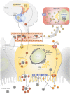
FIGURE 1
FIGURE 1
Scheme summarizing TH synthesis. TRH is produced by a specific group of neurons located in the paraventricular nucleus of the hypothalamus. When TRH reaches the pituitary gland, it binds to the TRH receptor expressed in the thyrotrophs, stimulating the expression and secretion of TSH into the circulation. TSH reaches the thyroid gland and binds to the TSHR located in the cell membrane of the thyroid follicles, setting in motion TH production in a process that includes the induction of iodine uptake by the NIS. Iodide is mobilized to the colloid via Pendrin action, and it is then oxidized by the thyroid peroxidase (TPO) using H2O2. Iodination of tyrosine residues forms monoiodotyrosines and diiodotyrosines that are coupled to form T3 and T4. T3 and T4 bound to TG are released in the colloid of the follicle. When needed, iodinated TG is catabolized in thyroid follicular epithelial cells to produce T3 and T4, which are then released into the circulation. In the bloodstream, THs can be found either free or bound to serum TH‐binding proteins (STHBP), such as thyroxine‐binding protein, transthyretin, and albumin. Free THs are able to enter into target cells in target tissues via membrane transporters. In target cells, deiodinases generate T3 from T4 by removing the iodine located at the 5´ position of T4. Intracellular T3 acts via genomic actions binding to the THR, where modulate gene expression, or via non‐genomic actions affecting signaling pathways such as integrin αvβ3 and PI3 K. THs also act via the nuclear THRβ in the hypothalamus and the pituitary to inhibit TRH and TSH production and secretion, completing a negative feedback loop that maintains physiological levels of THs. DIO: deiodinase. DUOX2: dual oxidase 2. DUOXA2: dual oxidase maturation factor 2. I: iodide. STHBP: serum TH‐binding proteins. Na+: sodium. NIS: sodium‐iodide symporter. T3: triiodothyronine. T4: thyroxine. Tg: thyroglobulin. TH: thyroid hormone. THR: thyroid hormone receptors. TPO: thyroid peroxidase. TRH: thyrotropin‐releasing hormone. TSH: thyrotropin. TSHR: thyrotropin receptor
FIGURE 2
FIGURE 2
Reference ranges and medical conditions associated with thyroid dysfunction. This figure defines reference ranges for primary hypothyroidism, secondary hypothyroidism, tertiary hypothyroidism, subclinical hypothyroidism, euthyroidism, subclinical hyperthyroidism, primary hyperthyroidism/thyroxine intoxication, and TSH‐producing adenoma/resistance to THs. TSH and T4 levels were defined as Hollowell et al. Total thyroxine can be converted from nM to µg/dl, dividing by 12.87. THs: thyroid hormones. TSH: thyrotropin
FIGURE 3
FIGURE 3
Scheme summarizing the processes regulated by THs in the main metabolic tissues. THs exert profound effects in metabolic tissues. THs enhance GK and MAFA expression in the pancreas favoring a rapid maturation and turnover of β cells. THs also potentiate insulin expression and secretion in the endocrine pancreas. Insulin‐target tissues respond increasing the activity of insulin signaling, which produces increased rates of lipolysis and gluconeogenesis in the liver and proteolysis and mitochondrial biogenesis in the skeletal muscle. Adipose tissues respond to THs increasing lipolysis and lipid mobilization. Browning/beiging of adipocytes occurs in the WAT and increasing thermogenesis via increased UCP expression and subsequent lipolysis occurs in the BAT. AKT, protein kinase B. FOXO: forkhead box O 1. GK: glucokinase. GLUT4: glucose transporter 4. MAFA: MAF bZIP transcription factor A

References

    1. Abe, E. , Marians, R. C. , Yu, W. , Wu, X.‐B. , Ando, T. , Li, Y. , Iqbal, J. , Eldeiry, L. , Rajendren, G. , Blair, H. C. , Davies, T. F. , & Zaidi, M. (2003). TSH is a negative regulator of skeletal remodeling. Cell, 115, 151–162. 10.1016/s0092-8674(03)00771-2 - DOI - PubMed
    1. Adami, H.‐O. , Chow, W.‐H. , Nyren, O. , Berne, C. , Linet, M. S. , Ekbom, A. , Wolk, A. , McLaughlin, J. K. , & Fraumeni, J. F. (1996). Excess risk of primary liver cancer in patients with diabetes mellitus. Journal of the National Cancer Institute, 88, 1472–1477. 10.1093/jnci/88.20.1472 - DOI - PubMed
    1. Aguayo‐Mazzucato, C. , Zavacki, A. M. , Marinelarena, A. , Hollister‐Lock, J. , El Khattabi, I. , Marsili, A. , Weir, G. C. , Sharma, A. , Larsen, P. R. , & Bonner‐Weir, S. (2013). Thyroid hormone promotes postnatal rat pancreatic beta‐cell development and glucose‐responsive insulin secretion through MAFA. Diabetes, 62, 1569–1580. 10.2337/db12-0849 - DOI - PMC - PubMed
    1. Aguayo‐Mazzucato, C. , Lee Jr, T. B. , Matzko, M. , Dilenno, A. , Rezanejad, H. , Ramadoss, P. , Scanlan, T. , Zavacki, A. M. , Larsen, P. R. , Hollenberg, A. , Colton, C. , Sharma, A. , & Bonner‐Weir, S. (2018). T3 induces both markers of maturation and aging in pancreatic beta‐cells. Diabetes, 67, 1322–1331. 10.2337/db18-0030 - DOI - PMC - PubMed
    1. Aguayo‐Mazzucato, C. , DiIenno, A. , Hollister‐Lock, J. , Cahill, C. , Sharma, A. , Weir, G. , Colton, C. , & Bonner‐Weir, S. (2015). MAFA and T3 drive maturation of both fetal human islets and insulin‐producing cells differentiated from hESC. The Journal of Clinical Endocrinology & Metabolism, 100(10), 3651–3659. 10.1210/jc.2015-2632 - DOI - PMC - PubMed

Publication types

Substances