. 2021 Dec;185(12):3593-3600.
doi: 10.1002/ajmg.a.61908. Epub 2020 Oct 13.

Novel pathogenic genomic variants leading to autosomal dominant and recessive Robinow syndrome

Affiliations

Novel pathogenic genomic variants leading to autosomal dominant and recessive Robinow syndrome

Chaofan Zhang et al. Am J Med Genet A. 2021 Dec.

Abstract

Robinow syndrome (RS) is a genetically heterogeneous disorder characterized by skeletal dysplasia and a distinctive facial appearance. Previous studies have revealed locus heterogeneity with rare variants in DVL1, DVL3, FZD2, NXN, ROR2, and WNT5A underlying the etiology of RS. The aforementioned "Robinow-associated genes" and their gene products all play a role in the WNT/planar cell polarity signaling pathway. We performed gene-targeted Sanger sequencing, exome sequencing, genome sequencing, and array comparative genomic hybridization on four subjects with a clinical diagnosis of RS who had not had prior DNA testing. Individuals in our cohort were found to carry pathogenic or likely pathogenic variants in three RS related genes: DVL1, ROR2, and NXN. One subject was found to have a nonsense variant (c.817C > T [p.Gln273*]) in NXN in trans with an ~1 Mb telomeric deletion on chromosome 17p containing NXN, which supports our contention that biallelic NXN variant alleles are responsible for a novel autosomal recessive RS locus. These findings provide increased understanding of the role of WNT signaling in skeletal development and maintenance. These data further support the hypothesis that dysregulation of the noncanonical WNT pathway in humans gives rise to RS.

Keywords: clinical diagnosis; deletion; missense; skeletal dysplasia; structural variant.


Conflict of interest statement

Disclosures

Baylor College of Medicine (BCM) and Miraca Holdings have formed a joint venture with shared ownership and governance of the Baylor Genetics (BG), which performs clinical microarray analysis and clinical exome sequencing. J.R.L. serves on the Scientific Advisory Board of the BG. J.R.L. has stock ownership in 23andMe, is a paid consultant for Regeneron Pharmaceuticals, and is a coinventor on multiple United States and European patents related to molecular diagnostics for inherited neuropathies, eye diseases, and bacterial genomic fingerprinting. The Department of Molecular and Human Genetics at Baylor College of Medicine derives revenue from molecular genetic and clinical genomics testing offered at BG. The other authors declare no competing financial interests.

Figures
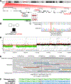
Figure 1.
Figure 1.. Location of DNA variants affecting NXN resulting in RRS
A: Below the chr17 ideogram, region p13.3 (red box) is expanded to show all genes in the genomic interval. The variant c.817C>T (red circle) is indicated on the NXN gene, which is a nonsense variant followed by a representation of subtelomeric deletion (green rectangle) containing NXN. This deletion does not include PAFAH1B1 or YWHAE related to the Miller-Diecker deletion syndrome. B: Family pedigree. Patient BAB10973 carries biallelic variants affecting NXN. Parental samples are not available for testing. C: Sanger sequencing results confirm the nonsense variant affecting NXN as detected by ES; D: Array CGH result (AMADID#085772) from chr17p reveals a telomeric deletion spanning 1.027 Mb that includes NXN (green probes shadowed by a red rectangle); E: Integrative Genomics Viewer (IGV) screenshot showing 30x GS Illumina short-reads spanning the breakpoint junction of the telomeric deletion. Grey dotted line indicates that the breakpoint junction maps at chr17:1,026,797. F: Sequencing alignment of the breakpoint junction from the telomeric deletion as confirmed by standard Sanger sequencing. The color-matched junction sequence is aligned to the telomeric repeats and the distal genomic references at ABR. Strand of alignment (+ or -) is indicated in parenthesis.
Figure 2.
Figure 2.. Robinow syndrome-associated genes involved in the WNT/PCP pathway identified in human subjects
The establishment of WNT/PCP signaling is vital for vertebrate development. All Robinow syndrome associated genes result in proteins that play a role in WNT/PCP signaling, therefore, dysregulation of the WNT pathway in humans may be the cause of Robinow syndrome. ROR2 is a co-receptor of FZD2, which binds to extracellular ligand WNT5A, together they can activate the non-canonical WNT signaling, which is associated with planar cell polarity in drosophila and the equivalent convergent-extension movement during gastrulation in vertebrates (Isao Oishi1, 2003; Sokol, 2000; Yamanaka et al., 2002). DVLs are key downstream mediators of the WNT pathway and NXN was found to be a negative regulator of the WNT pathway. This model is adapted and revised from a previous version (White et al., 2018). We removed a potential candidate gene RAC3 since the clinical phenotype of the patient who has a potentially pathogenic variant in RAC3 is more consistent with clinically distinct RAC3-related neurodevelopmental disorder (Costain et al., 2019; de Curtis, 2019; Hiraide et al., 2019) than with DRS.

References

    1. Abyzov A, Urban AE, Snyder M, & Gerstein M (2011). CNVnator: an approach to discover, genotype, and characterize typical and atypical CNVs from family and population genome sequencing. Genome Research, 21(6), 974–984. doi:10.1101/gr.114876.110 - DOI - PMC - PubMed
    1. Ali BR, Jeffery S, Patel N, Tinworth LE, Meguid N, Patton MA, & Afzal AR (2007). Novel Robinow syndrome causing mutations in the proximal region of the frizzled-like domain of ROR2 are retained in the endoplasmic reticulum. Human Genetics, 122(3–4), 389–395. doi:10.1007/s00439-007-0409-0 - DOI - PubMed
    1. Beck CR, Carvalho CMB, Akdemir ZC, Sedlazeck FJ, Song X, Meng Q, . . . Lupski JR (2019). Megabase Length Hypermutation Accompanies Human Structural Variation at 17p11.2. Cell, 176(6), 1310–1324 e1310. doi:10.1016/j.cell.2019.01.045 - DOI - PMC - PubMed
    1. Ben-Shachar S, Khajavi M, Withers MA, Shaw CA, van Bokhoven H, Brunner HG, & Lupski JR (2009). Dominant versus recessive traits conveyed by allelic mutations - to what extent is nonsense-mediated decay involved? Clinical Genetics, 75(4), 394–400. doi:10.1111/j.1399-0004.2008.01114.x - DOI - PubMed
    1. Bi W, Sapir T, Shchelochkov OA, Zhang F, Withers MA, Hunter JV, . . . Reiner O (2009). Increased LIS1 expression affects human and mouse brain development. Nature Genetics, 41(2), 168–177. doi:10.1038/ng.302 - DOI - PMC - PubMed

Publication types

MeSH terms

Supplementary concepts