. 2020 Nov-Dec;22(10):598-607.
doi: 10.1016/j.micinf.2020.10.004. Epub 2020 Oct 10.

Non-synonymous mutations of SARS-CoV-2 leads epitope loss and segregates its variants

Affiliations

Non-synonymous mutations of SARS-CoV-2 leads epitope loss and segregates its variants

Aayatti Mallick Gupta et al. Microbes Infect. 2020 Nov-Dec.

Abstract

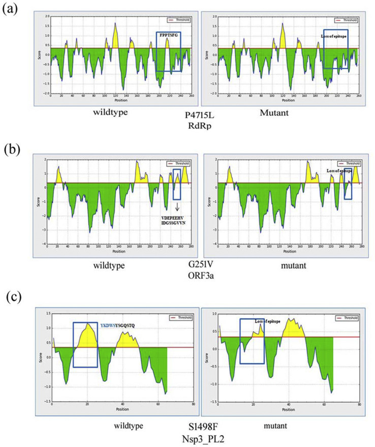
The non-synonymous mutations of SARS-CoV-2 isolated from across the world have been identified during the last few months. The surface glycoprotein spike of SARS-CoV-2 forms the most important hotspot for amino acid alterations followed by the ORF1a/ORF1ab poly-proteins. It is evident that the D614G mutation in spike glycoprotein and P4715L in RdRp is the important determinant of SARS-CoV-2 evolution since its emergence. P4715L in RdRp, G251V in ORF3a and S1498F of Nsp3 is associated with the epitope loss that may influence pathogenesis caused by antibody escape variants. The phylogenomics distinguished the ancestral viral samples from China and most part of Asia, isolated since the initial outbreak and the later evolved variants isolated from Europe and Americas. The evolved variants have been found to predominant globally with the loss of epitopes from its proteins. These have implications for SARS-CoV-2 transmission, pathogenesis and immune interventions.

Keywords: COVID-19; Epitope loss; Non-synonymous mutation; Phylogenomics; SARS-CoV-2.


Conflict of interest statement

Declaration of competing interest The authors wish to declare that they do not have any conflict of interest.

Figures

Fig. 1
Fig. 1
(a) Structure of the SARS-CoV-2 genome. (b) The relative occurrence of non-synonymous mutation across the genome of SARS-CoV-2. Black indicates neutral mutation and red are deleterious mutations.
Fig. 2
Fig. 2
Epitope loss linked with non-synonymous mutations. The predicted score above the threshold level is the Yellow region showing epitope. (a) Effect of P4715L mutation in RdRp. (a) B-cell epitope in non-mutated RdRp (left) and P4715L mutant (right). It is linked with such epitope loss FPPTSFG from the site. (b) Epitope loss linked with ORF3a G251V. The B-cell epitope of wildtype ORF3a (left). G251V mutant (right) causes loss of DGSSGVV(250 …. 256aa). (c) Epitope loss linked with S1498F in the papain-like protease domain of NSP3 in the ORF1a region. The B-cell epitope of wildtype sample (left), S1498F mutation causes the loss YKDWS (right).
Fig. 3
Fig. 3
P4715L RdRp and D614G spike variants show co-occurrence. (a) The incidence (%) of occurrence of P4715 and L4715 of RdRp in various geographical locations. (b) The incidence (%) of occurrence of D614 and G614 of spike protein in various geographical locations. (c) The overlap in the mutation across the world is depicted from the combined plot. The distribution of these two unique mutant sites of vital SARS-CoV-2 proteins can be completely superimposed.
Fig. 4
Fig. 4
Phylogenomics with different SARS-CoV-2 viral samples across the world. The tree is distinctly divided into 2 clades: green shows the original (ancestral) form of the virus isolated from Dec, 2019 to Feb, 2020 and red clades are the evolved variant isolated after Feb, 2020 to April, 2020. Loss of epitope is found in the evolved variant due to non-synonymous mutation. Indian isolates are highlighted in purple belonging to both the green and red clades. The samples from Kerala, India are Wuhan representative (within green clade) and the rest are the variants from Italy (within red clade).

References

    1. Poon L.L.M., Peiris M. Emergence of a novel human coronavirus threatening human health. Nat Med. 2020;26:317–319. - PMC - PubMed
    1. Chan J.F., Yuan S., Kok K.H., To K.K.W., Chu H., Yang J. A familial cluster of pneumonia associated with the 2019 novel coronavirus indicating person-to person transmission: a study of a family cluster. Lancet. 2020;395:514–523. - PMC - PubMed
    1. Chen N., Zhou M., Dong X., Qu J., Gong F., Han Y. Epidemiological and clinical characteristics of 99 cases of 2019 novel coronavirus pneumonia in Wuhan, China: a descriptive study. Lancet. 2020;395:507–513. - PMC - PubMed
    1. Cucinotta D., Vanelli M. WHO declares COVID-19 a pandemic. Acta Biomed. 2020;91:157–160. - PMC - PubMed
    1. Zhou P., Yang X.L., Wang X.G., Hu B., Zhang L., Zhang W. A pneumonia outbreak associated with a new coronavirus of probable bat origin. Nature. 2020;579:270–273. - PMC - PubMed

MeSH terms