. 2020 Oct 8;12(10):3068.
doi: 10.3390/nu12103068.

Oligo-Fucoidan Improves Diabetes-Induced Renal Fibrosis via Activation of Sirt-1, GLP-1R, and Nrf2/HO-1: An In Vitro and In Vivo Study

Affiliations

Oligo-Fucoidan Improves Diabetes-Induced Renal Fibrosis via Activation of Sirt-1, GLP-1R, and Nrf2/HO-1: An In Vitro and In Vivo Study

Wen-Chun Yu et al. Nutrients. .

Abstract

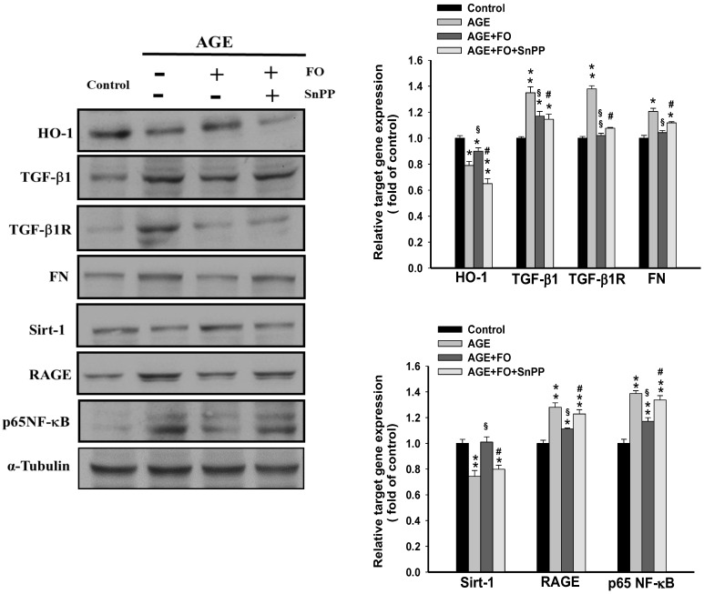
Fucoidan extracted from brown algae has multiple beneficial functions. In this study, we investigated the effects of low-molecular-weight fucoidan (oligo-FO) on renal fibrosis under in vitro and in vivo diabetic conditions, and its molecular mechanisms. Advanced glycation product (AGE)-stimulated rat renal proximal tubular epithelial cells (NRK-52E) and diabetic mice induced by high-fat diet and intraperitoneal injection of streptozotocin and nicotinamide were used. Oligo-FO treatment significantly inhibited anti-high mobility group box 1 (HMGB1)/RAGE/ anti-nuclear factor-kappa B (NF-κB)/transforming growth factor-β1 (TGF-β1)/TGF-β1R/Smad 2/3/fibronectin signaling pathway and HIF-1α activation in AGE-stimulated NRK-52E cells. Conversely, the expression and activity of Sirt-1; the levels of ubiquitin-specific peptidase 22 (USP22), p-AMPK, glucagon-like peptide-1 receptor (GLP-1R), and heme oxygenase-1 (HO-1); and Nrf2 activation were remarkably increased by oligo-FO in AGE-stimulated cells. However, the above effects of oligo-FO were greatly diminished by inhibiting Sirt-1, HO-1, or GLP-1R activity. Similar changes of these pro-fibrotic genes in the kidney and a marked attenuation of renal injury and dysfunction were observed in oligo-FO-treated diabetic mice. These findings indicated that the inhibitory effects of the oligo-FO on diabetes-evoked renal fibrosis are mediated by suppressing TGF-β1-activated pro-fibrogenic processes via Sirt-1, HO-1, and GLP-1R dependence. Collectively, fucoidan-containing foods or supplements may be potential agents for ameliorating renal diseases due to excessive fibrosis.

Keywords: Sirt-1; diabetes; fucoidan; glucagon-like peptide-1 receptor; renal fibrosis; transforming growth factor-β.


Conflict of interest statement

The authors declare no conflict of interest.

Figures

Figure 1
Figure 1
The effects of fucoidan on Sirt-1 activity, anti-high mobility group box 1 (HMGB1) cellular location, and fibrosis-related signaling pathways. The rat renal proximal tubular epithelial cells (NRK-52E) cells were incubated with advanced glycation product (AGE) (100 μg/mL) for 24 h followed by treatment with fucoidan (100 μg/mL) for 24 h in the presence or absence of EX527 (200 nM). The Sirt-1 activity (A), the expression of fibrosis-related genes (B), the cytosolic and nuclear levels of HMGB1 (C), and the association of Sirt-1 with HMGB1 in the nucleus (D) were determined in various groups. FO: fucoidan. Results were expressed as the mean ± SEM (n = 5). * p < 0.05, ** p < 0.01 vs. control group (untreated NRK-52E cells); § p < 0.05 vs. AGE-treated alone cells; # p < 0.05 vs. AGE and fucoidan-treated cells.
Figure 2
Figure 2
The effects of fucoidan on Nrf2 activation and the expression of glucagon-like peptide-1 receptor (GLP-1R), AMP-activated protein kinase (AMPK), and heme oxygenase-1(HO-1). The NRK-52E cells were incubated with AGE (100 μg/mL) for 24 h followed by treatment with fucoidan (100 μg/mL) for 24 h in the presence or absence of EX527 (200 nM). The expression of glucagon-like peptide-1 receptor (GLP-1R), AMP-activated protein kinase (AMPK), p-AMPK, Kelch-like ECH-associated protein 1 (Keap1), and HO-1 (A), and the nuclear level of Nrf2 (B) was determined in various groups. Results were expressed as the mean ± S.E.M (n = 5). * p < 0.05, ** p < 0.01 vs. control group; § p < 0.05, §§ p < 0.01 vs. AGE-treated alone cells; # p < 0.05, ## p < 0.01 vs. AGE and fucoidan-treated cells.
Figure 3
Figure 3
The effects of tin protoporphyrin IX (SnPP) on fucoidan-regulated target gene expression. The NRK-52E cells were incubated with AGE (100 μg/mL) for 24 h, followed by treatment with fucoidan (100 μg/mL) for 24 h in the presence or absence of SnPP (20 μM). The alterations of target gene expression were determined in various groups. Results were expressed as the mean ± S.E.M (n = 5). * p < 0.05, ** p < 0.01 vs. control group; § p < 0.05, §§ p < 0.01 vs. AGE-treated alone cells; # p < 0.05 vs. AGE and fucoidan-treated cells.
Figure 4
Figure 4
The effects of exendin-3 (9-39) (ED9-39) on fucoidan-regulated target gene expression. The NRK-52E cells were incubated with AGE (100 μg/mL) for 24 h followed by treatment with fucoidan (100 μg/mL) for 24 h in the presence or absence of ED9-39 (1 mM). The protein expression of target genes (A), and the nuclear level of hypoxia-inducible factor-1α (HIF-1α) (B), were determined in various groups. Results were expressed as the mean ± S.E.M (n = 5). * p < 0.05, ** p < 0.01 vs. control group; § p < 0.05, §§ p < 0.01 vs. AGE-treated alone cells; # P < 0.05 vs. AGE and fucoidan-treated cells.
Figure 5
Figure 5
The effects of fucoidan on fibrosis-related gene expression, Sirt-1 activity, and the histological changes in the kidney of diabetic nephropathy mice. The expression of target genes (A), the Sirt-1 activity (B), and the histological changes evaluated by Masson staining and PAS staning (C) in the kidney of the diabetic mice were examined in different groups. Results were expressed as the mean ± SEM (n = 8). * p < 0.05, ** p < 0.01, *** p < 0.001 vs. normal mice; # p < 0.05, ## p < 0.01 vs. untreated diabetic nephropathy mice.
Figure 6
Figure 6
The effects of fucoidan on urine levels of BUN, creatinine, and albumin, in diabetic nephropathy mice. After treatment with fucoidan (1) (300 mg/kg, p.o.) for 6 weeks, the urine levels of BUN, creatinine, and albumin were examined in different groups (A). Results were expressed as the mean ± SEM (n = 8). * p < 0.05, ** p < 0.01 vs. normal mice; # p < 0.05, ## p < 0.01 vs. untreated diabetic mice. The proposed schematic diagram of the protective effect of fucoidan against diabetic nephropathy (B).

Similar articles

Cited by

References

    1. Kanwar Y.S., Sun L., Xie P., Liu F.-Y., Chen S. A glimpse of various pathogenetic mechanisms of diabetic nephropathy. Annu. Rev. Pathol. Mech. Dis. 2011;6:395–423. doi: 10.1146/annurev.pathol.4.110807.092150. - DOI - PMC - PubMed
    1. Fioretto P., Mauer M. Histopathology of diabetic nephropathy. Semin. Nephrol. 2007;27:195–207. doi: 10.1016/j.semnephrol.2007.01.012. - DOI - PMC - PubMed
    1. Meng X.M., Tang P.M., Li J., Lan H.Y. TGF-beta/Smad signaling in renal fibrosis. Front. Physiol. 2015;6:82. doi: 10.3389/fphys.2015.00082. - DOI - PMC - PubMed
    1. Svensson M., Sundkvist G., Arnqvist H.J., Björk E., Blohmé G., Bolinder J., Henricsson M., Nyström L., Torffvit O., Waernbaum I., et al. Signs of nephropathy may occur early in young adults with diabetes despite modern diabetes management: Results from the nationwide population-based Diabetes Incidence Study in Sweden (DISS) Diabetes Care. 2003;26:2903–2909. doi: 10.2337/diacare.26.10.2903. - DOI - PubMed
    1. Rahman S., Islam R. Mammalian Sirt1: Insights on its biological functions. Cell Commun. Signal. 2011;9:11. doi: 10.1186/1478-811X-9-11. - DOI - PMC - PubMed

MeSH terms