Review
. 2020 Oct 8;9(10):2255.
doi: 10.3390/cells9102255.

Syncytia in Fungi

Affiliations
Review

Syncytia in Fungi

Alexander P Mela et al. Cells. .

Abstract

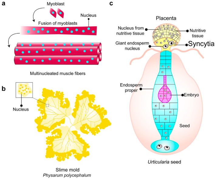
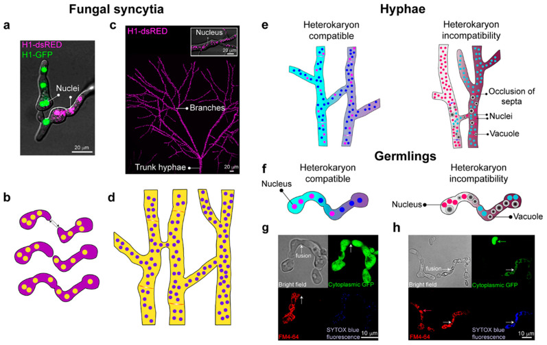
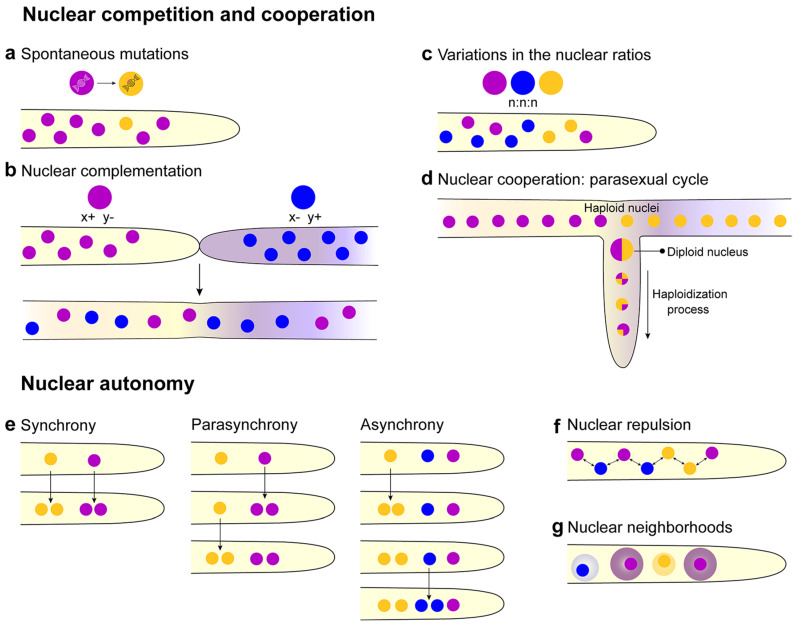
Filamentous fungi typically grow as interconnected multinucleate syncytia that can be microscopic to many hectares in size. Mechanistic details and rules that govern the formation and function of these multinucleate syncytia are largely unexplored, including details on syncytial morphology and the regulatory controls of cellular and molecular processes. Recent discoveries have revealed various adaptations that enable fungal syncytia to accomplish coordinated behaviors, including cell growth, nuclear division, secretion, communication, and adaptation of the hyphal network for mixing nuclear and cytoplasmic organelles. In this review, we highlight recent studies using advanced technologies to define rules that govern organizing principles of hyphal and colony differentiation, including various aspects of nuclear and mitochondrial cooperation versus competition. We place these findings into context with previous foundational literature and present still unanswered questions on mechanistic aspects, function, and morphological diversity of fungal syncytia across the fungal kingdom.

Keywords: filamentous fungi; heterokaryon; morphology; nucleus; syncytia.


Conflict of interest statement

The authors declare no conflict of interest.

Figures

Figure 1
Figure 1
Examples of syncytia in nature. (a) Myoblasts are fused to form multinucleated muscle fibers. (b) Slime mold Physarum polycephalum, multinucleate protoplasm formed by the fusion of individual amoebae. (c) Endospermal–placental syncytia in developmental stages of Urticularia seeds, the formation of syncytia occurs in the placenta. The syncytia harbor two populations of nuclei, a nucleus from the nutritive tissue, and a giant endosperm nucleus (modified from [8]).
Figure 2
Figure 2
Formation of interconnected fungal syncytia. (a) The fusion of compatible strains of Neurospora crassa, whose nuclei are labeled with either histone H1-GFP or H1-dsRED (green and magenta, respectively). (b) Genetically identical germs grow to each other (dashed arrows), fuse, and give rise to interconnected multinucleate syncytia. (c) Heterogeneity of the mycelial network and syncytia formation. Magenta dots are nuclei marked with histone H1-DsRed. The box shows a close-up of a hypha, showing the marked nuclei. (d) Hyphal fusion within a colony contributes to an interconnected syncytium. (e) In hyphae, heterokaryon formation can occur when there are no differences at het (heterokaryon) or vic (vegetative incompatibility) loci. In contrast, genetic differences at these loci result in heterokaryon incompatibility, which triggers compartmentalization of the fusion compartment due to occlusion of the septum, vacuolization of the hyphae, and eventual cell death. (f) In germlings, heterokaryon formation can occur when there are no differences at the rcd-1 (regulator of cell death-1) and plp-1/sec-9 (patatin-like phospholipase-1) loci. In contrast, differences at these loci result in heterokaryon incompatibility, rapidly triggering a cell death reaction that is a similar process in hyphae [38,39]. (g) Micrographs show the fusion of compatible germlings. One of the germlings is marked with cytoplasmic Green Fluorescent Protein (GFP) (green) and has undergone cell fusion with a compatible germling stained with FM4-64 fluorescent dye (red). Fusion is evident by the fact that GFP fluorescence can be observed in both germlings due to cytoplasmic mixing, and cell death does not occur, as indicated with the absence of SYTOX Blue fluorescence (death cell stain). (h) Cell fusion between germlings with genetic differences at the plp/sec-9 loci results in rapid cellular vacuolization and death, as demonstrated with the staining of SYTOX Blue fluorescence. White arrows indicate fusion events. Micrographs also show two germlings that have not undergone cell fusion and are healthy (green; GFP and red arrows; FM4-64). Micrographs (g,h) courtesy of Dr. Jens Heller (UCB Glass Laboratory).
Figure 3
Figure 3
Nuclear patterns in multinucleate syncytia. (a) Spontaneous mutation in nuclei can give that nucleus an advantage or disadvantage over the rest of the nuclei in the syncytial population. (b) Nuclear complementation can occur if a nucleus lacks a gene (x− or y−) encoding a function necessary for survival. x− or y− function can be complemented by the presence of a second nucleus, which is functional for that gene (x+ or y+). Complementation between nuclei in a syncytium can occur with spontaneous mutations (a) or via complementation upon fusion with another individual that can produce the missing component. (c) In multinucleate syncytia, variations in the nuclear ratios can occur, where one nucleus can dominate. (d) Generation of nuclear heterogeneity through the parasexual cycle. Haploid nuclei in a heterokaryon, formed either by spontaneous mutation or via fusion with a different strain, undergo karyogamy to form a heterozygous diploid nucleus. Repeated rounds of mitotic recombination and mitotic nondisjunction result in loss of chromosomes, producing haploid nuclei with unique genotypes. (e) Different patterns of nuclei division in syncytia. Synchrony: all the nuclei divide at the same time. Parasynchrony, the mitosis is initiated in one nucleus, and then linearly, the adjacent nucleus starts to divide after the first one. Asynchrony, the nuclei divide independently of each other (modified from [93]. (f) The nuclei show repulsion to delimit their cytoplasmic territory [96], and within a hypha, a regular number of nuclei per unit volume of cytoplasm (#N/C) is observed [102]. (g) Nuclear neighborhoods can be organized around nuclei that affect cell cycle regulation and, potentially, other regulatory processes (modified from [111]).

References

    1. Zeldovich V.B., Clausen C.H., Bradford E., Fletcher D.A., Maltepe E., Robbins J.R., Bakardjiev A.I. Placental syncytium forms a biophysical barrier against pathogen invasion. PLoS Pathog. 2013;9:e1003821. doi: 10.1371/journal.ppat.1003821. - DOI - PMC - PubMed
    1. Abmayr S.M., Zhuang S., Geisbrecht E.R. Myoblast fusion in Drosophila. Methods Mol. Biol. 2008;475:75–97. - PubMed
    1. Scheven B.A., Haas E.W.K.-D., Wassenaar A.M., Nijweide P.J. Differentiation kinetics of osteoclasts in the periosteum of embryonic bones in vivo and in vitro. Anat. Rec. 1986;214:418–423. doi: 10.1002/ar.1092140413. - DOI - PubMed
    1. Daniel J.W., Jarlfors U. Plasmodial ultrastructure of the myxomycete Physarum polycephalum. Tissue Cell. 1972;4:15–36. doi: 10.1016/S0040-8166(72)80003-X. - DOI - PubMed
    1. Guttes E., Guttes S., Rusch H.P. Morphological observations on growth and differentation of Physarum polycephalum grown in pure culture. Dev. Biol. 1961;3:588–614. doi: 10.1016/0012-1606(61)90034-3. - DOI - PubMed

Publication types