Review
. 2020 Oct 9;10(10):1429.
doi: 10.3390/biom10101429.

Role of Glutathione in Cancer: From Mechanisms to Therapies

Affiliations
Review

Role of Glutathione in Cancer: From Mechanisms to Therapies

Luke Kennedy et al. Biomolecules. .

Abstract

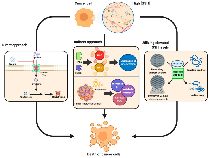
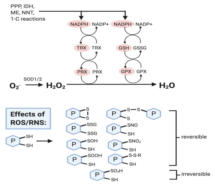
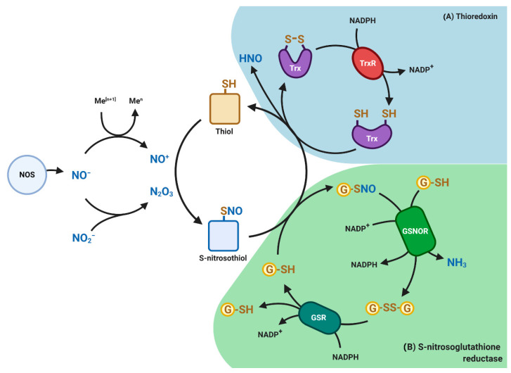
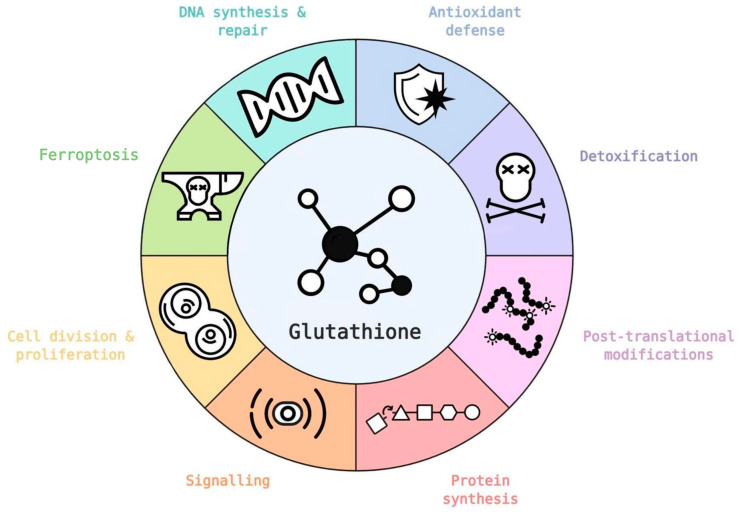
Glutathione (GSH) is the most abundant non-protein thiol present at millimolar concentrations in mammalian tissues. As an important intracellular antioxidant, it acts as a regulator of cellular redox state protecting cells from damage caused by lipid peroxides, reactive oxygen and nitrogen species, and xenobiotics. Recent studies have highlighted the importance of GSH in key signal transduction reactions as a controller of cell differentiation, proliferation, apoptosis, ferroptosis and immune function. Molecular changes in the GSH antioxidant system and disturbances in GSH homeostasis have been implicated in tumor initiation, progression, and treatment response. Hence, GSH has both protective and pathogenic roles. Although in healthy cells it is crucial for the removal and detoxification of carcinogens, elevated GSH levels in tumor cells are associated with tumor progression and increased resistance to chemotherapeutic drugs. Recently, several novel therapies have been developed to target the GSH antioxidant system in tumors as a means for increased response and decreased drug resistance. In this comprehensive review we explore mechanisms of GSH functionalities and different therapeutic approaches that either target GSH directly, indirectly or use GSH-based prodrugs. Consideration is also given to the computational methods used to describe GSH related processes for in silico testing of treatment effects.

Keywords: S-nitrosation; ferroptosis; glutathione; metabolism modeling; nitrosoglutathione; reactive oxygen species; tumor metabolism; tumor therapy.


Conflict of interest statement

The authors declare no conflict of interest.

Figures

Figure 1
Figure 1
Different approaches for cancer therapy related to glutathione functions. High levels of glutathione (GSH) observed in the majority of tumors combined with its role in tumorigenesis, inflammation response and tumor microenvironment properties are explored for different therapy approaches. Direct approaches are aimed at blocking synthesis of GSH leading to ferroptosis of cells or more efficient chemotherapy; indirect methods target inflammation response or tumor microenvironment in order to make tumors more susceptible to the response of the immune system as well as immunotherapies; elevated levels of GSH and possibly other active molecules in tumor cells are used in the prodrug approach, where the drug is only activated after entering tumor cells utilizing GSH in the activation reaction or neutralizing it following activation with other factors.
Figure 2
Figure 2
Reactive oxygen species (ROS) removal and effects. NADPH levels are crucial in ROS removal mechanisms and is produced through the pentose phosphate pathway (PPP), isocitrate dehydrogenase (IDH), malic enzyme (ME), nicotinamide nucleotide transhydrogenase (NNT), and one-carbon reactions (e.g., tetrahydrofolate metabolism). NADPH reduces oxidized forms of thioredoxin (TRX) and glutathione (GSSG), and these reduced forms (shown in red) then activate peroxiredoxin (PRX) and glutathione peroxidase (GPX) so that they can reduce H2O2 to water (H2O). ROS (mainly H2O2) and RNS (e.g., nitric oxide) can cause the oxidative modification of protein cysteine thiol residues, most of which are reversible, and serve to protect them from oxidative damage, and to modify protein activity. Refer to text for details.
Figure 3
Figure 3
Redox-based post-translational modifications mediated by nitric oxide (NO). Nitric oxide synthases (NOS) generates NO gas which reacts with thiol-containing molecules such as proteins and glutathione (GSH) to form S-nitrosothiols or S-nitrosoglutatione (GSNO), respectively. Shown are (A) Thioredoxin (Trx) pathway (blue) and (B) S-nitrosoglutathione reductase (GSNOR) pathway (green). GSNOR and TR catalyzes the denitrosation of protein nitrosothiols and GSNO. By reducing GSNO to GSSG and NH3, GSNOR indirectly controls the levels of S-nitrosothiols and associated signal transduction. Glutathione (GSSG) is reduced back to GSH by glutathione reductase (GSR) at the expense of NADPH. Similarly, Trx mediates denitrosation of protein nitrosothiols by releasing nitroxyl (HNO) group to form reduced thioredoxin at the expense of NADPH. HNO can diffuse and react in the surrounding environment. An imbalance in NO signaling can promote tumorigenesis and tumor progression.
Figure 4
Figure 4
Glutathione has a number of critical roles in healthy and tumor cells, in both cases supporting cell metabolism and survival. Several glutathione functionalities are directly or indirectly regulated by oncogenes (icons based on database [50]). Refer to text for further details.
Figure 5
Figure 5
Biosynthesis pathway of glutathione (GSH) from amino acid precursors. GSH is synthesized in two consecutive ATP-dependent reactions within the cytosol coupled with GSH degradation to its constituent amino acids via 5-oxoproline and cysteinyl glycine, which forms the GSH cycle. Precursor sources are indicated by green arrows. GSH synthesis is regulated through inhibition of GCLC by GSH. GCLC, glutathione cysteine ligase (GCL) catalytic subunit; GCLM, glutathione cysteine ligase (GCL) modifier subunit; GS, glutathione synthase. Further explanation of synthesis processes is provided in the text.
Figure 6
Figure 6
Relative GSH and GSSG concentrations vary slightly across cancer cell lines (data presented by Li et al., [65] and obtained from https://portals.broadinstitute.org/ccle). Figure shows concentration of GSSG as a function of the concentration of GSH in the same cell line (concentrations shown in mM). Insert plot shows differences in the ratio of GSH/GSSG across 928 cell lines.
Figure 7
Figure 7
Induction of metabolic reprogramming in recipient cells by internalization of producer cell-derived exosomes. Producer cells (e.g., cancer-associated fibroblasts) contain high levels of tricarboxylic acid (TCA) cycle metabolites, amino acids and lipids that can fuel the metabolic activity of recipient tumor cells contributing to tumor progression. Producer cells (e.g., glioma cells) derived exosomes can also harbor mRNA encoding ribosomal, oxidative phosphorylation and glycolytic proteins that can reprogram glycolysis and oxidative phosphorylation in recipient cells resulting in oncogenic reprogramming and transformation. Intraexosomal cargoes, such as metabolites, amino acids and mRNA are based on the studies of Cuperlovic-Culf et al., [156] Zhao et al., [149] and Zheng et al., [150]. EV, extracellular vesicles, such as exosomes; MVB, multivesicular body; OXPHOS, oxidative phosphorylation. Refer to text for more details.

References

    1. Reth M. Hydrogen peroxide as second messenger in lymphocyte activation. Nat. Immunol. 2002;3:1129–1134. doi: 10.1038/ni1202-1129. - DOI - PubMed
    1. Bienert G.P., Schjoerring J.K., Jahn T.P. Membrane transport of hydrogen peroxide. Biochim. Biophys. Acta. 2006;1758:994–1003. doi: 10.1016/j.bbamem.2006.02.015. - DOI - PubMed
    1. Hauck A.K., Bernlohr D.A. Oxidative stress and lipotoxicity. J. Lipid Res. 2016;57:1976–1986. doi: 10.1194/jlr.R066597. - DOI - PMC - PubMed
    1. Davies M.J. Protein oxidation and peroxidation. Biochem. J. 2016;473:805–825. doi: 10.1042/BJ20151227. - DOI - PMC - PubMed
    1. Moldogazieva N.T., Mokhosoev I.M., Feldman N.B., Lutsenko S.V. ROS and RNS signalling: Adaptive redox switches through oxidative/nitrosative protein modifications. Free Radic. Res. 2018;52:507–543. doi: 10.1080/10715762.2018.1457217. - DOI - PubMed

Publication types