Review
. 2020 Oct 9;9(10):964.
doi: 10.3390/antiox9100964.

The Impact of Mitochondrial Deficiencies in Neuromuscular Diseases

Affiliations
Review

The Impact of Mitochondrial Deficiencies in Neuromuscular Diseases

Judith Cantó-Santos et al. Antioxidants (Basel). .

Abstract

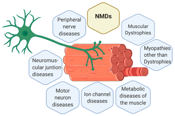
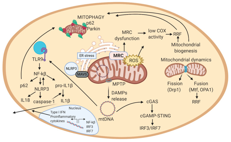
Neuromuscular diseases (NMDs) are a heterogeneous group of acquired or inherited rare disorders caused by injury or dysfunction of the anterior horn cells of the spinal cord (lower motor neurons), peripheral nerves, neuromuscular junctions, or skeletal muscles leading to muscle weakness and waste. Unfortunately, most of them entail serious or even fatal consequences. The prevalence rates among NMDs range between 1 and 10 per 100,000 population, but their rarity and diversity pose difficulties for healthcare and research. Some molecular hallmarks are being explored to elucidate the mechanisms triggering disease, to set the path for further advances. In fact, in the present review we outline the metabolic alterations of NMDs, mainly focusing on the role of mitochondria. The aim of the review is to discuss the mechanisms underlying energy production, oxidative stress generation, cell signaling, autophagy, and inflammation triggered or conditioned by the mitochondria. Briefly, increased levels of inflammation have been linked to reactive oxygen species (ROS) accumulation, which is key in mitochondrial genomic instability and mitochondrial respiratory chain (MRC) dysfunction. ROS burst, impaired autophagy, and increased inflammation are observed in many NMDs. Increasing knowledge of the etiology of NMDs will help to develop better diagnosis and treatments, eventually reducing the health and economic burden of NMDs for patients and healthcare systems.

Keywords: damage-associated molecular patterns (DAMPs); mitochondrial respiratory chain (MRC); mitophagy; neuromuscular diseases (NMDs); oxidative stress; reactive oxygen species (ROS).


Conflict of interest statement

The authors declare no conflict of interest. The funders had no role in the design of the study; in the collection, analyses, or interpretation of data; in the writing of the manuscript, or in the decision to publish the results.

Figures

Figure 1
Figure 1
Representation of a motor unit according to the classification of the main neuromuscular diseases (NMDs). Muscle cells and neurons interact in the neuromuscular junction (NMJ) where muscle cells receive the instructions from the lower motor neurons to perform the actions required. Thus, body movement is actioned by muscle fibers but controlled by the nervous system. Consequently, alterations in muscle fibers or nerves can both trigger a NMD. Figure created with BioRender.com.
Figure 2
Figure 2
Schematic view of the mechanistic model of mitochondrial-related pathways in a cell. 1. MRC generates ATP in the inner mitochondrial membrane. In this process, also ROS are produced. 2. In case of excessive ROS levels, mitochondrial membrane potential decreases, allowing MPTP to open and release DAMPs (mtDNA, cardiolipin, ceramides) to the cytosol. These DAMPs can trigger an inflammatory response. In this scheme, the inflammatory cascade represented is the one activated by mtDNA. mtDNA activates cGAS in cytosol, that eventually induces the expression of interferon-related genes (IRF3/IRF7). In parallel, mtDNA activates TLR9, which activates NF-kB, and thus, NLRP3 inflammasome, expression of pro-inflammatory cytokines, and among them, IL-1 response. 3. Increased ROS levels can result from MRC dysfunction. To compensate dysfunctional mitochondria, mitochondrial biogenesis increases the number of mitochondria in a cell. Excessive mitochondria around muscle cells form RRF. 4. Excessive ROS, RRF, inflammation, and ER stress damage mitochondria, which alterations are sensed by autophagy and mitophagy. Autophagy and mitophagy remove damaged mitochondria, and consequently stop the inflammatory response, supporting repair mechanisms in the cell. If autophagy and mitophagy are altered, mitochondria cannot be recycled, leading to apoptosis. Abbreviations: MRC: mitochondrial respiratory chain; ROS: reactive oxygen species; MPTP: mitochondrial permeability transition pore; DAMPs: damage-associated molecular patterns; mtDNA: mitochondrial DNA; cGAS: cyclic GMP-AMP synthase; TLR9: toll-like receptor 9; NF-kB: nuclear factor kappa-light-chain-enhancer of activated B cells; NLRP3: NOD-like receptor protein 3; RRF: ragged-red fibers; ER: endoplasmic reticulum; MAVS: mitochondrial antiviral signaling protein. Figure created with BioRender.com.

Similar articles

Cited by

References

    1. Diagnosis—European Reference Network—EURO-NMD. [(accessed on 22 April 2020)]; Available online: https://ern-euro-nmd.eu/
    1. Scotton C., Passarelli C., Neri M., Ferlini A. Biomarkers in rare neuromuscular diseases. Exp. Cell Res. 2013;325:44–49. doi: 10.1016/j.yexcr.2013.12.020. - DOI - PubMed
    1. Bonne G., Rivier F., Hamroun D. The 2018 version of the gene table of monogenic neuromuscular disorders (nuclear genome) Neuromuscul. Disord. 2017;27:1152–1183. doi: 10.1016/j.nmd.2017.10.005. - DOI - PubMed
    1. Deenen C.W.J., Horlings G.C.C., Verschuuren J.G.M.J., Verbeek L.M.A., van Engelen G.M.B. The Epidemiology of Neuromuscular Disorders: A Comprehensive Overview of the Literature. J. Neuromuscul. Dis. 2015;2:73–85. doi: 10.3233/JND-140045. - DOI - PubMed
    1. Van Putten M., Hmeljak J., Aartsma-Rus A., Dowling J.J. Moving neuromuscular disorders research forward: From novel models to clinical studies. Dis. Model. Mech. 2020;13:44370. doi: 10.1242/dmm.044370. - DOI - PMC - PubMed