Review
. 2020 Oct 9;9(10):2263.
doi: 10.3390/cells9102263.

Neuropathic Itch

Affiliations
Review

Neuropathic Itch

James Meixiong et al. Cells. .

Abstract

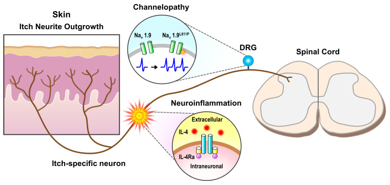
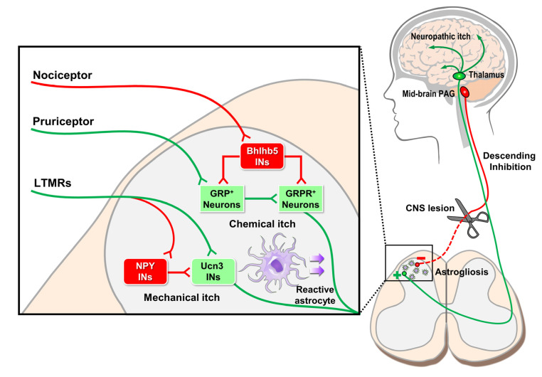
Neurologic insults as varied as inflammation, stroke, and fibromyalgia elicit neuropathic pain and itch. Noxious sensation results when aberrantly increased afferent signaling reaches percept-forming cortical neurons and can occur due to increased sensory signaling, decreased inhibitory signaling, or a combination of both processes. To treat these symptoms, detailed knowledge of sensory transmission, from innervated end organ to cortex, is required. Molecular, genetic, and behavioral dissection of itch in animals and patients has improved understanding of the receptors, cells, and circuits involved. In this review, we will discuss neuropathic itch with a focus on the itch-specific circuit.

Keywords: inflammation; neuropathy; pruritus.


Conflict of interest statement

The authors declare no conflict of interest.

Figures

Figure 1
Figure 1
Neuropathic itch of the peripheral nervous system. Small diameter itch-sensing nerves of the dorsal root ganglia (DRG) ramify in skin and synapse with secondary neurons in the outer laminar layers of the dorsal horn of the spinal cord. Various insults along this peripheral neuronal tract can result in neuropathic itch. These include increased neurite outgrowth in diseased skin, neuroinflammatory activation via IL-4/IL-4Ra receptor, and genetic channelopathies such as Nav1.9L811P which has been associated with increased itch nerve activity.
Figure 2
Figure 2
Models of coding for itch and pain. Different models of coding have been proposed to explain the relationship between itch and pain. (a) In the labeled-line coding model, the itch-specific sensory neuron (left) and pain-specific neuron (right) each constitutes a dedicated pathway that only responds to corresponding pruritogens (i.e., chloroquine) or algogens (i.e., capsaicin). (b) In the intensity coding model, the itch and pain pathways are comprised of the same population of sensory neurons and circuits. Low-to medium intensity stimuli evoke low firing rates of the sensory neurons and result in itch sensation (left). On the contrary, high-intensity stimuli evoke robust firing rates of the same sensory neurons and result in pain sensation (right). (c) In the spatial contrast coding model, focal stimulation of the individual nociceptors induces itch sensation (left). Contrarily, co-stimulation of the same population of sensory neurons with the neighboring nociceptors evokes the pain sensation (right).
Figure 3
Figure 3
Neuropathic itch of the central nervous system. Peripheral itch nerves synapse with secondary Gastrin-releasing peptide (GRP)+ itch neurons of the spinal cord which then activate tertiary GRP receptor (GRPR)+ neurons. GRPR+ neurons decussate anteriorly to form the spinothalamic tract and synapse with thalamic neurons which communicate with various brain regions such as the sensory cortex and insula to produce itch sensation. In the spinal cord, numerous interneuron (INs) populations control itch processing. For example, nociceptor activation of Bhlhb5 INs inhibits GRP+ and GRPR+ neuron signaling of chemical itch. Similarly, peripheral low-threshold mechanoreceptors (LTMRs) can both enhance mechanical itch by activating excitatory Ucn3 INs and inhibit mechanical itch via inhibitory NPY INs. In addition to these peripheral inputs, higher-level brain regions, such as the mid-brain periaqueductal gray (PAG), provide descending inhibitory feedback onto spinal cord itch neurons. If descending inhibitory pathways are disrupted, then itch becomes disinhibited, and neuropathic itch results. Finally, chronic itch conditions can cause reactive spinal astrogliosis, a non-neuronal mechanism whereby itch signaling is enhanced.

References

    1. Oaklander A.L. Common neuropathic itch syndromes. Acta Derm. Venereol. 2012;92:118–125. doi: 10.2340/00015555-1318. - DOI - PubMed
    1. International Association for the Study of Pain Home Page. [(accessed on 20 September 2020)]; Available online: https://www.iasp-pain.org/GlobalYear/NeuropathicPain.
    1. Oaklander A.L. Neuropathic Itch. In: Carstens E., Akiyama T., editors. Itch: Mechanisms and Treatment. 1st ed. CRC Press; Boca Raton, FL, USA: 2014.
    1. Cheng X.Q., Liang X.Z., Wei S., Ding X., Han G.H., Liu P., Sun X., Quan Q., Tang H., Zhao Q., et al. Protein microarray analysis of cytokine expression changes in distal stumps after sciatic nerve transection. Neural Regen Res. 2020;15:503–511. doi: 10.4103/1673-5374.266062. - DOI - PMC - PubMed
    1. Steinhoff M., Oaklander A.L., Szabo I.L., Stander S., Schmelz M. Neuropathic itch. Pain. 2019;160:S11–S16. doi: 10.1097/j.pain.0000000000001551. - DOI - PubMed