. 2020 Oct 9;8(10):403.
doi: 10.3390/biomedicines8100403.

Stearic Acid and TNF-α Co-Operatively Potentiate MIP-1α Production in Monocytic Cells via MyD88 Independent TLR4/TBK/IRF3 Signaling Pathway

Affiliations

Stearic Acid and TNF-α Co-Operatively Potentiate MIP-1α Production in Monocytic Cells via MyD88 Independent TLR4/TBK/IRF3 Signaling Pathway

Shihab Kochumon et al. Biomedicines. .

Abstract

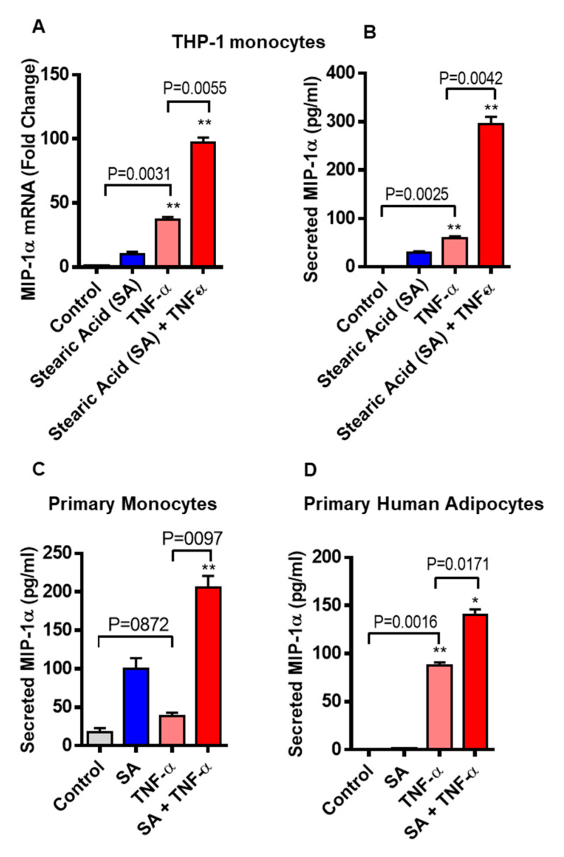
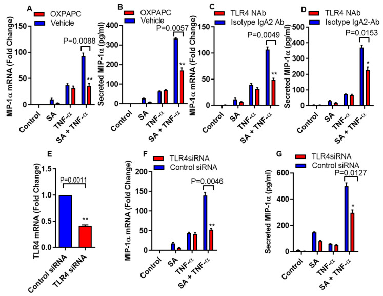
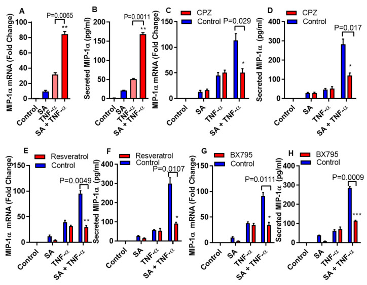
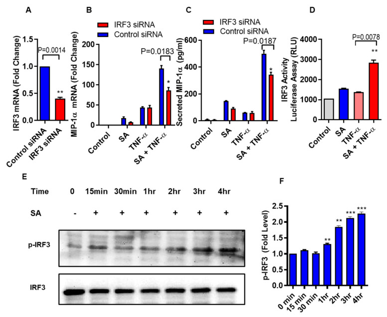
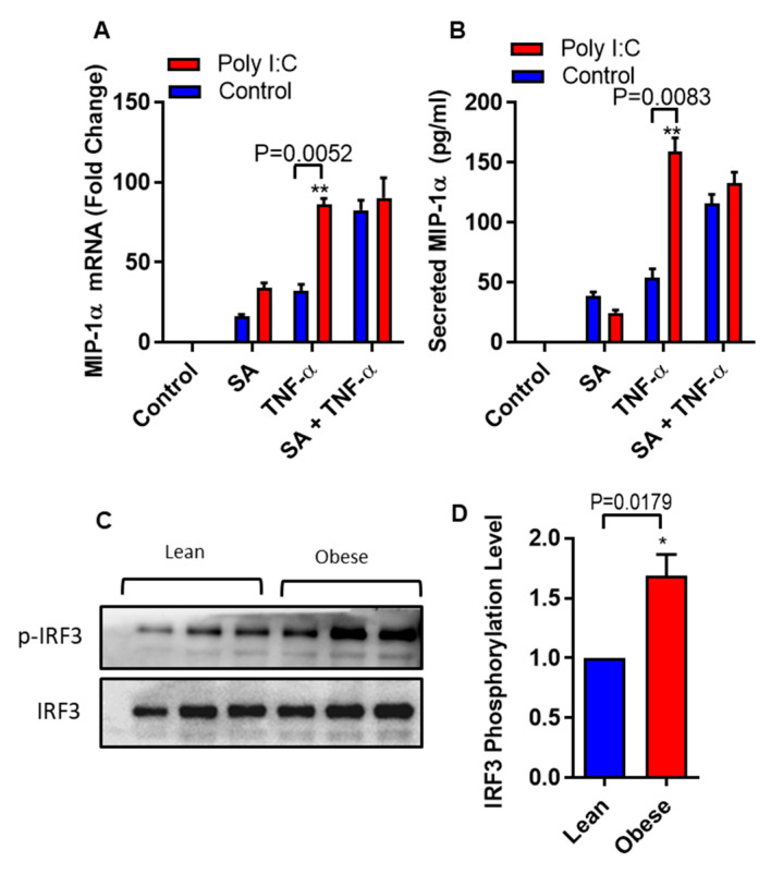
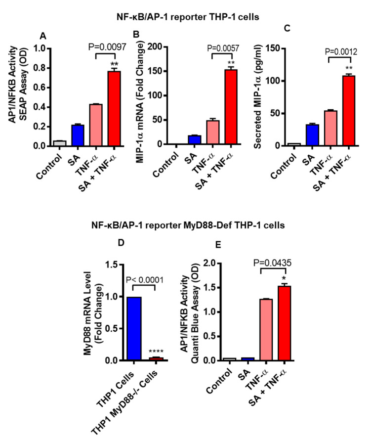
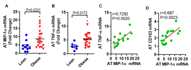
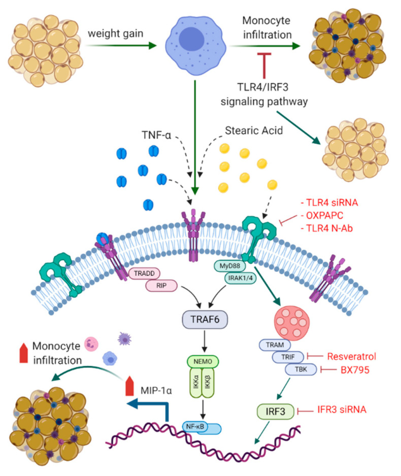
Increased circulatory and adipose tissue expression of macrophage inflammatory protein (MIP)-1α (CC motif chemokine ligand-3/CCL3) and its association with inflammation in the state of obesity is well documented. Since obesity is associated with increases in both stearic acid and tumor necrosis factor α (TNF-α) in circulation, we investigated whether stearic acid and TNF-α together could regulate MIP-1α/CCL3 expression in human monocytic cells, and if so, which signaling pathways were involved in MIP-1α/CCL3 modulation. Monocytic cells were treated with stearic acid and TNF-α resulted in enhanced production of MIP-1α/CCL3 compared to stearic acid or TNF-α alone. To explore the underlying mechanisms, cooperative effect of stearic acid for MIP-α/CCL3 expression was reduced by TLR4 blocking, and unexpectedly we found that the synergistic production of MIP-α/CCL3 in MyD88 knockout (KO) cells was not suppressed. In contrast, this MIP-α/CCL3 expression was attenuated by inhibiting TBK1/IRF3 activity. Cells deficient in IRF3 did not show cooperative effect of stearate/TNF-α on MIP-1α/CCL3 production. Furthermore, activation of IRF3 by polyinosinic-polycytidylic acid (poly I:C) produced a cooperative effect with TNF-α for MIP-1α/CCL3 production that was comparable to stearic acid. Individuals with obesity show high IRF3 expression in monocytes as compared to lean individuals. Furthermore, elevated levels of MIP-1α/CCL3 positively correlate with TNF-α and CD163 in fat tissues from individuals with obesity. Taken together, this study provides a novel model for the pathologic role of stearic acid to produce MIP-1α/CCL3 in the presence of TNF-α associated with obesity settings.

Keywords: IRF3; MIP-1α; TLR4; TNF-α/CCL3; TRIF/TBK; obesity; stearic acid.


Conflict of interest statement

The authors declare no conflict of interest.

Figures

Figure 1
Figure 1
Co-treatment of stearic acid and tumor necrosis factor α (TNF-α) enhances macrophage inflammatory protein (MIP)-1α (CC motif chemokine ligand-3/CCL3) production. (A) THP-1 cells were incubated with stearic acid (200 µM) and TNFα (10 ng/mL) for 24 h. Cells and culture media were collected. Total RNA was extracted from the cells and CCL3 mRNA was quantified by real time PCR. Relative mRNA expression was expressed as fold change (B) Secreted MIP-1α/CCL3 protein in culture media was determined by ELISA. (C,D) Primary human monocytes and adipocytes were treated with stearic acid and TNF-α for 24 h. MIP-1α/CCL3 mRNA and secreted protein were determined. The results obtained from minimum three independent experiments with three replicates of each experiment are shown. All data are expressed as mean ± SEM (n ≥ 3); * P < 0.05; ** P < 0.01, when compared with stearic acid or TNF-α alone.
Figure 2
Figure 2
Disruption of TLR4 suppresses cooperative effect of stearic acid with TNF-α for MIP-1α/CCL3 production. (A,B) THP-1 cells were incubated with oxidized 1-palmitoyl-2-arachidonyl-sn- glycero-3-phosphorylcholine (OXPAPC) (30 µg/mL) for 1 h and then treated with stearic acid and TNF-α for 24 h. Cells and culture media were collected. MIP-1α/CCL3 gene expression was determined by real time PCR and secreted MIP-1α/CCL3 protein was determined in culture media by ELISA. (C,D). Monocytic cells were treated with 2 µg/mL of neutralizing TLR4 mAb or isotype-matched control (IgA2) for 40 min. Antibody-treated cells were treated with stearic acid and TNF-α for 24 h. MIP-1α/CCL3 mRNA and secreted MIP-1α/CCL3 protein were determined. (EG) Monocytic cells were transfected with either control or TLR4 siRNA. TLR4-deficient cells were stimulated with stearic acid and TNF-α for 24 h. Knockdown efficiency of transfection was checked by TLR4 gene expression with qRT-PCR. MIP-1α/CCL3 mRNA and secreted MIP-1α/CCL3 protein were determined. The results obtained from minimum three independent experiments with three replicates of each experiment are shown. All data are expressed as mean ± SEM (n ≥ 3); * P < 0.05; ** P < 0.01when compared with Stearic acid or TNF-α alone.
Figure 3
Figure 3
Stearic acid cooperative effect on MIP-1α/CCL3 is MyD88 independent and dependent on TRIF/TBK1. (A,B) Cells deficient in MyD88 activity (THP-1 defMyD cells) were treated with stearic acid (200 µM), 0.1% BSA (control) or TNF-α (10 ng/mL) alone or in combination. Cells and culture media were collected after 24 h. CCL3 mRNA and secreted MIP-1α/CCL3 protein were determined. (C,D) Monocytic cells were pretreated with chlorpromazine (CPZ; an inhibitor of endocytosis; 10 µM) for 1 h and then treated as indicated for 24 h. MIP-1α/CCL3 mRNA and secreted MIP-1α/CCL3 protein were determined. (E,F) Cells were treated with resveratrol (a TRIF inhibitor; 5 uM) for 30 min followed by treatments as indicated. MIP-α expression was determined. (G,H) Cells were incubated with BX795 (an inhibitor for TBK1/IKKε; 100 nM) for 1 h and then treated for 24 h as indicated. MIP-1α/CCL3 expression was determined. The results obtained from minimum three independent experiments with three replicates of each experiment are shown. All data are expressed as mean ± SEM (n ≥ 3); * P < 0.05; ** P < 0.01; *** P < 0.001, when compared with stearic acid or TNF-α alone.
Figure 4
Figure 4
Stearic acid cooperative effect with TNF-α for MIP-1α/CCL3 production requires IRF3. (A) THP-1 monocytic cells were transfected with either control or IRF3 siRNA and incubated for 36 h. Real time PCR was done to measure IRF3 expression. (B,C) IRF3 deficient THP-1 cells were stimulated with stearic acid and TNF-α. MIP-1α/CCL3 expression was determined. The results obtained from minimum three independent experiments with three replicates of each experiment are shown. (D) IRF3 activity reporter monocytic cells were treated with stearic acid (200 µM) or 0.1% BSA (control) or TNF-α (10 ng/mL) or in combination. Culture media were collected after 24 h. Cell culture media were assayed for luciferase activity representing the degree of IRF3/ISRE activation using Quanti-Luc medium. (E) Western blot analysis showed that stearic acid induced IRF3 phosphorylation in a time dependent manner in THP-1 monocytes, verifies the role of IRF3 in the cooperative effect of stearic acid in the TNF-α mediated production of MIP-1α/CCL3. (F) Expression of phosphorylated IRF3 is shown as determined by densitometry of western blot bands. * P < 0.05; ** P < 0.01; *** P < 0.001.
Figure 5
Figure 5
Polyinosinic-polycytidylic acid (poly I:C) act as a substitute of stearic acid cooperative effect on MIP-1α/CCL3 production. (A,B) THP-1 cells were treated (via transfection) with poly I:C (5 µg) for 2 h and then incubated with BSA (control) or stearic acid or TNF-α for 24 h. MIP-1α/CCL3 mRNA and protein were determined. The results obtained from minimum three independent experiments with three replicates of each experiment are shown. All data are expressed as mean ± SEM (n ≥ 3); * P < 0.05; ** P < 0.01, when compared with stearic acid or TNF-α alone. (C,D) Western blot analysis showed that individuals with obesity have significant higher levels of phospho-IRF3 in monocytes compared to lean individuals (P = 0.0179).
Figure 6
Figure 6
THP-1-XBlue cells (THP-1 cells stably expressing a secreted embryonic alkaline phosphatase (SEAP) reporter inducible by NF-κB and AP-1) were treated with stearic acid or BSA or TNF-α for 24 h. Culture media were collected. Cell culture media were assayed for SEAP reporter activity (degree of NF-κB/AP-1 activation) (A), along with MIP-1α gene and protein expression (B,C). Next, THP-1-XBlue™-defMyD cells (Cells deficient in MyD88 activity) were used showing defective gene expression of MyD88 (D). These cells were also treated with stearic acid (200 μM), TNF-α (10 ng/mL) and 0.1% BSA for 24 h and SEAP reporter activity (degree of NF-κB/AP-1 activation) is shown (E). The results obtained from three independent experiments are shown. The data are presented as mean ± SD. * P < 0.05; ** P < 0.01; **** P < 0.001, when compared with stearic acid or TNF-α alone.
Figure 7
Figure 7
MIP-1α/CCL3 is associated with TNF-α in subcutaneous fat from obese humans. The level of (A) MIP-1α/CCL3 (8.67 ± 1.22 folds, p = 0.0221) and (B) TNF-α (5.09 ± 0.48 folds, p = 0.0172) mRNA levels were significantly higher in obese as compared to lean individuals (4.28 ± 0.99 folds for MIP-1 α, and 2.72 ± 0.54 folds for TNF-α). In obese adipose tissue, a strong positive correlation was found between (C) MIP-1α/CCL3 and TNF-α mRNA expression (r = 0.7292; p = 0.0022), and (D) MIP-1α/CCL3 and CD163 (r = 0.686, p = 0.0023).
Figure 8
Figure 8
Schematic illustration of signaling pathways underlying the cooperative relationship between stearic acid and TNF-α for MIP-1α/CCL3 production. Blocking TLR4/IRF3 signaling pathways significantly suppress the cooperative production of MIP-1α/CCL3 by stearic acid/TNF-α. TLR: Toll like receptor; TNF-α: Tumor necrosis factor α; MyD88: Myeloid differentiation factor 88; IRAK: Interleukin-1 receptor-associated kinase; TRIF: TIR domain–containing adapter-inducing IFN-β; IRF3: Interferon regulatory factor-3; TBK: TANK-binding kinase 1; TRAM: Toll-receptor-associated molecule; TRAF6: tumor-necrosis factor receptor-associated factor 6: IKK-Iκ-B kinase; NF-kB: Nuclear factor-κB; TRADD: Tumor necrosis factor receptor 1-associated death domain protein; RIP: receptor-interacting protein; NEMO: NF-kappa-B essential modulator. BioRender.com was used for Figure 8.

References

    1. Yaribeygi H., Farrokhi F.R., Butler A.E., Sahebkar A. Insulin resistance: Review of the underlying molecular mechanisms. J. Cell. Physiol. 2019;234:8152–8161. doi: 10.1002/jcp.27603. - DOI - PubMed
    1. Choe S.S., Huh J.Y., Hwang I.J., Kim J.I., Kim J.B. Adipose tissue remodeling: Its role in energy metabolism and metabolic disorders. Front. Endocrinol. 2016;7:30. doi: 10.3389/fendo.2016.00030. - DOI - PMC - PubMed
    1. Lee B.C., Lee J. Cellular and molecular players in adipose tissue inflammation in the development of obesity-induced insulin resistance. Biochim. Biophys. Acta. 2014;1842:446–462. doi: 10.1016/j.bbadis.2013.05.017. - DOI - PMC - PubMed
    1. Gao D., Madi M., Ding C., Fok M., Steele T., Ford C., Hunter L., Bing C. Interleukin-1beta mediates macrophage-induced impairment of insulin signaling in human primary adipocytes. Am. J. Physiol. Endocrinol. Metab. 2014;307:E289–E304. doi: 10.1152/ajpendo.00430.2013. - DOI - PMC - PubMed
    1. Stephens J.M., Pekala P.H. Transcriptional repression of the c/ebp-alpha and glut4 genes in 3t3-l1 adipocytes by tumor necrosis factor-alpha. Regulations is coordinate and independent of protein synthesis. J. Biol. Chem. 1992;267:13580–13584. - PubMed