Review
. 2020 Oct 9;21(20):7452.
doi: 10.3390/ijms21207452.

The Dual Role of Glutamatergic Neurotransmission in Alzheimer's Disease: From Pathophysiology to Pharmacotherapy

Affiliations
Review

The Dual Role of Glutamatergic Neurotransmission in Alzheimer's Disease: From Pathophysiology to Pharmacotherapy

Vidyasagar Naik Bukke et al. Int J Mol Sci. .

Abstract

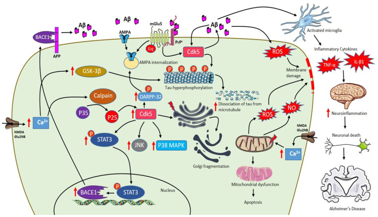
Alzheimer's disease (AD) is an age-related dementia and neurodegenerative disorder, characterized by Aβ and tau protein deposition impairing learning, memory and suppressing synaptic plasticity of neurons. Increasing evidence suggests that there is a link between the glucose and glutamate alterations with age that down-regulates glucose utilization reducing glutamate levels in AD patients. Deviations in brain energy metabolism reinforce the development of AD by hampering glutamate levels in the brain. Glutamate is a nonessential amino acid and the major excitatory neurotransmitter synthesized from glucose. Alterations in cerebral glucose and glutamate levels precede the deposition of Aβ plaques. In the brain, over 40% of neuronal synapses are glutamatergic and disturbances in glutamatergic function have been implicated in pathophysiology of AD. Nevertheless, targeting the glutamatergic system seems to be a promising strategy to develop novel, improved therapeutics for AD. Here, we review data supporting the involvement of the glutamatergic system in AD pathophysiology as well as the efficacy of glutamatergic agents in this neurodegenerative disorder. We also discuss exciting new prospects for the development of improved therapeutics for this devastating disorder.

Keywords: AD; AMPA; EAAT1/2; NMDA; ageing; amyoid-β; glucose; glutamate; metabotropic receptors; tau; therapeutic targets.


Conflict of interest statement

The authors declare that the research was conducted in the absence of any commercial or financial relationships that could be construed as a potential conflict of interest.

Figures

Figure 1
Figure 1
Presynaptic terminals release glutamate activating ionotropic receptors on postsynaptic neurons. In normal condition, N-methyl-d-aspartate (NMDA) NR2A activation induces increase in calcium levels favouring induction of long-term potentiation (LTP) through metabolic pathways (extracellular signal-related protein kinase (ERK), CaMK II, cyclic adenosine monophosphate response element-binding protein (CREB)). Excess glutamate left is taken up by astrocytes through EAAT2 converting into glutamine and glutamate by glutamine synthetase and glutaminase respectively (black arrows). The synthesized glutamate is transported into vesicles by VGlut1/2. Conversely in Alzheimer’s disease (AD), Aβ oligomers interfere with NMDA receptors increasing the spillover of glutamate (red arrows) to extrasynaptic sites activating NMDA NR2B receptors increasing excess calcium levels inhibiting prosurvival pathways. Imbalance in glutamate/glutamine cycle (black-dashed arrows) is also reported in the figure.
Figure 2
Figure 2
Schematic representation of AD pathophysiology involving Aβ, extrasynaptic NMDA Glu2NB, mGlu5 and role of activated microglia. Activation of NMDA Glu2NB receptors increases calcium levels inducing p35 to p25 cleavage mediated by calpain and p25 is activates cdk-5 enhancing DARPP-32 phosphorylation eventually leading to AMPA receptor internalization. Increase in Aβ levels activates Cdk-5 that plays a major role in Golgi fragmentation and tau phosphorylation leading to dissociation of tau from microtubules. Furthermore, Cdk-5 mediates phosphorylation of signal transducer and activator of transcription 3 (STAT3), which increases β-site APP cleaving enzyme 1 (BACE1) transcription resulting in an increase of Aβ content. Moreover, Aβ-PrPc-mGlu5 combination leads to AMPA internalization. Excess increase in levels of Ca2+ in mitochondria results in the generation of reactive oxygen species (ROS) and NO, inhibition of ATP synthesis, mitochondrial permeability transition pore (mPTP) opening, release of cytochrome c, activation of caspases and leading to apoptosis. In addition, Aβ also initiates a spectrum of neuroinflammation by activating microglia that plays a detrimental role in the expression of pro-inflammatory cytokines like interleukins and tumor necrosis factor-α (TNF-α) influencing neurodegeneration. (red arrows indicate “increase”).

References

    1. Querfurth H.W., LaFerla F.M. Alzheimer’s disease. N. Engl. J. Med. 2010;362:329–344. doi: 10.1056/NEJMra0909142. - DOI - PubMed
    1. Chen G.F., Xu T.H., Yan Y., Zhou Y.R., Jiang Y., Melcher K., Xu H.E. Amyloid beta: Structure, biology and structure-based therapeutic development. Acta Pharmacol. Sin. 2017;38:1205–1235. doi: 10.1038/aps.2017.28. - DOI - PMC - PubMed
    1. Coronel R., Palmer C., Bernabeu-Zornoza A., Monteagudo M., Rosca A., Zambrano A., Liste I. Physiological effects of amyloid precursor protein and its derivatives on neural stem cell biology and signaling pathways involved. Neural Regen. Res. 2019;14:1661–1671. doi: 10.4103/1673-5374.257511. - DOI - PMC - PubMed
    1. Larson M.E., Lesne S.E. Soluble Abeta oligomer production and toxicity. J. Neurochem. 2012;120:125–139. doi: 10.1111/j.1471-4159.2011.07478.x. - DOI - PMC - PubMed
    1. Rogaeva E., Meng Y., Lee J.H., Gu Y., Kawarai T., Zou F., Katayama T., Baldwin C.T., Cheng R., Hasegawa H., et al. The neuronal sortilin-related receptor SORL1 is genetically associated with Alzheimer disease. Nat. Genet. 2007;39:168–177. doi: 10.1038/ng1943. - DOI - PMC - PubMed