

Splicing Characteristics of Dystrophin Pseudoexons and Identification of a Novel Pathogenic Intronic Variant in the DMD Gene
- PMID: 33050418
- PMCID: PMC7650627
- DOI: 10.3390/genes11101180
Splicing Characteristics of Dystrophin Pseudoexons and Identification of a Novel Pathogenic Intronic Variant in the DMD Gene
Abstract
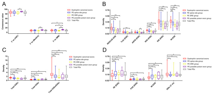
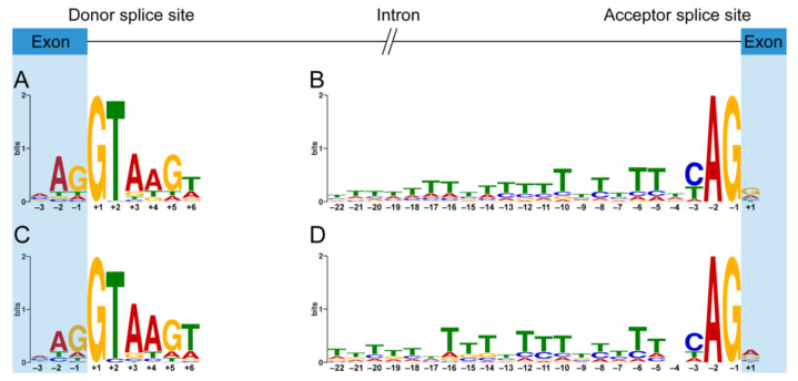
Pseudoexon (PE) inclusion has been implicated in various dystrophinopathies; however, its splicing characteristics have not been fully investigated. This study aims to analyze the splicing characteristics of dystrophin PEs and compare them with those of dystrophin canonical exons (CEs). Forty-two reported dystrophin PEs were divided into a splice site (ss) group and a splicing regulatory element (SRE) group. Five dystrophin PEs with characteristics of poison exons were identified and categorized as the possible poison exon group. The comparative analysis of each essential splicing signal among different groups of dystrophin PEs and dystrophin CEs revealed that the possible poison exon group had a stronger 3' ss compared to any other group. As for auxiliary SREs, different groups of dystrophin PEs were found to have a smaller density of diverse types of exonic splicing enhancers and a higher density of several types of exonic splicing silencers compared to dystrophin CEs. In addition, the possible poison exon group had a smaller density of 3' ss intronic splicing silencers compared to dystrophin CEs. To our knowledge, our findings indicate for the first time that poison exons might exist in DMD (the dystrophin gene) and present with different splicing characteristics than other dystrophin PEs and CEs.
Keywords: DMD; canonical exon; intronic variants; pseudoexon; splicing characteristics.
Conflict of interest statement
L.T. and C.Z. are employees of Running Gene Inc. Both of them analyzed the Sanger sequencing data. The other authors declare no conflicts of interest.
Figures
Similar articles
-
Pseudoexons of the DMD Gene.J Neuromuscul Dis. 2020;7(2):77-95. doi: 10.3233/JND-190431. J Neuromuscul Dis. 2020. PMID: 32176650 Free PMC article. Review.
-
DMD pseudoexon mutations: splicing efficiency, phenotype, and potential therapy.Ann Neurol. 2008 Jan;63(1):81-9. doi: 10.1002/ana.21290. Ann Neurol. 2008. PMID: 18059005
-
When a mid-intronic variation of DMD gene creates an ESE site.Neuromuscul Disord. 2014 Dec;24(12):1111-7. doi: 10.1016/j.nmd.2014.07.003. Epub 2014 Aug 1. Neuromuscul Disord. 2014. PMID: 25193336
-
Categorization of 77 dystrophin exons into 5 groups by a decision tree using indexes of splicing regulatory factors as decision markers.BMC Genet. 2012 Mar 31;13:23. doi: 10.1186/1471-2156-13-23. BMC Genet. 2012. PMID: 22462762 Free PMC article.
-
Exon skipping therapy for Duchenne muscular dystrophy.Adv Drug Deliv Rev. 2015 Jun 29;87:104-7. doi: 10.1016/j.addr.2015.05.008. Epub 2015 May 14. Adv Drug Deliv Rev. 2015. PMID: 25980936 Review.
Cited by
-
Role of Splicing Regulatory Elements and In Silico Tools Usage in the Identification of Deep Intronic Splicing Variants in Hereditary Breast/Ovarian Cancer Genes.Cancers (Basel). 2021 Jul 3;13(13):3341. doi: 10.3390/cancers13133341. Cancers (Basel). 2021. PMID: 34283047 Free PMC article.
-
A novel deep intronic variant introduce dystrophin pseudoexon in Becker muscular dystrophy: A case report.Heliyon. 2024 Mar 19;10(6):e28020. doi: 10.1016/j.heliyon.2024.e28020. eCollection 2024 Mar 30. Heliyon. 2024. PMID: 38545205 Free PMC article.
-
Tissue- and cell-specific whole-transcriptome meta-analysis from brain and retina reveals differential expression of dystrophin complexes and new dystrophin spliced isoforms.Hum Mol Genet. 2023 Jan 27;32(4):659-676. doi: 10.1093/hmg/ddac236. Hum Mol Genet. 2023. PMID: 36130212 Free PMC article.
-
Whole-Genome Sequencing Identified New Structural Variations in the DMD Gene That Cause Duchenne Muscular Dystrophy in Two Girls.Int J Mol Sci. 2023 Sep 1;24(17):13567. doi: 10.3390/ijms241713567. Int J Mol Sci. 2023. PMID: 37686372 Free PMC article.
-
30 Years Since the Proposal of Exon Skipping Therapy for Duchenne Muscular Dystrophy and the Future of Pseudoexon Skipping.Int J Mol Sci. 2025 Feb 3;26(3):1303. doi: 10.3390/ijms26031303. Int J Mol Sci. 2025. PMID: 39941071 Free PMC article. Review.
References
-
- Bladen C.L., Salgado D., Monges S., Foncuberta M.E., Kekou K., Kosma K., Dawkins H., Lamont L., Roy A.J., Chamova T., et al. The TREAT-NMD DMD global database: Analysis of more than 7000 duchenne muscular dystrophy mutations. Hum. Mutat. 2015;36:395–402. doi: 10.1002/humu.22758. - DOI - PMC - PubMed
Publication types
MeSH terms
Substances
LinkOut - more resources
Full Text Sources
Research Materials
