. 2020 Oct 11;21(20):7486.
doi: 10.3390/ijms21207486.

Synergic Therapeutic Potential of PEA-Um Treatment and NAAA Enzyme Silencing In the Management of Neuroinflammation

Affiliations

Synergic Therapeutic Potential of PEA-Um Treatment and NAAA Enzyme Silencing In the Management of Neuroinflammation

Giovanna Casili et al. Int J Mol Sci. .

Abstract

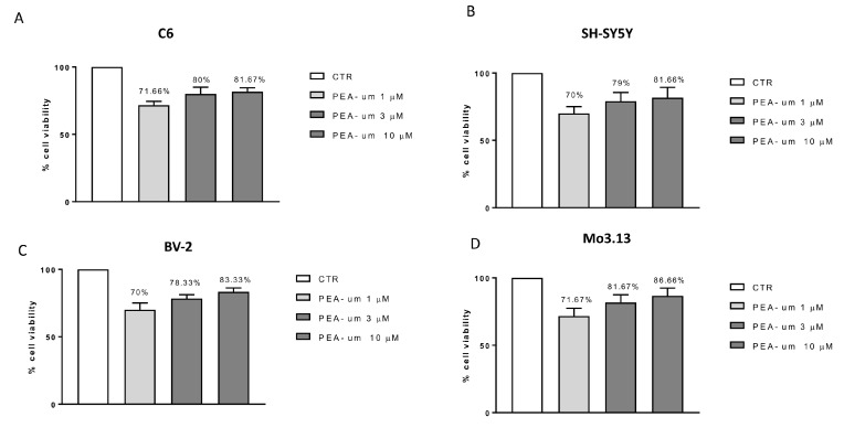
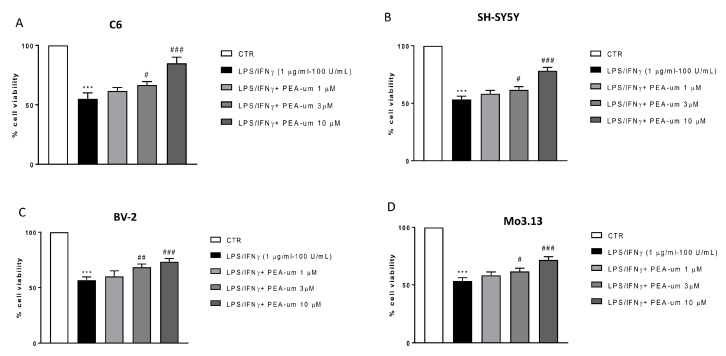
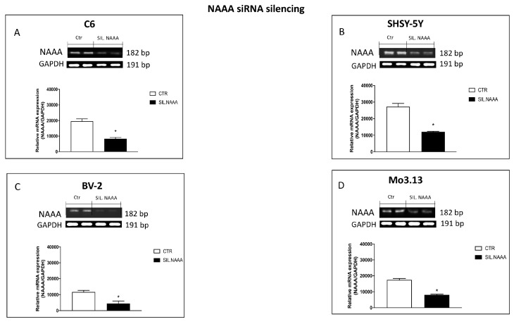
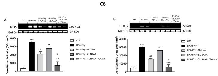
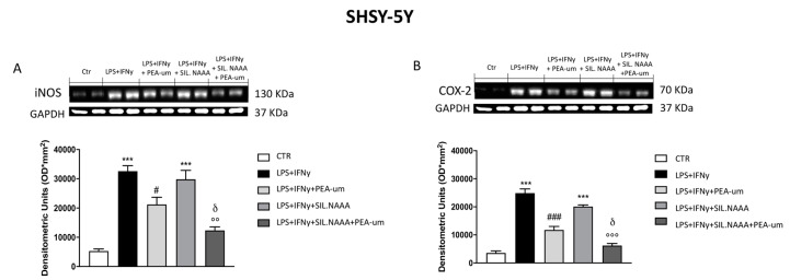
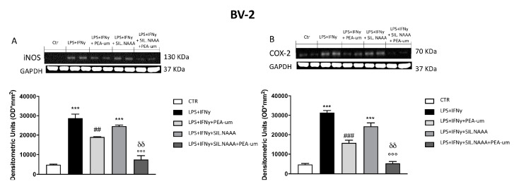
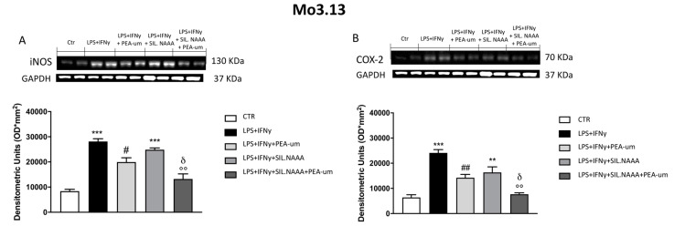
Inflammation is a key element in the pathobiology of neurodegenerative diseases and sees the involvement of different neuronal and non-neuronal cells as players able to respond to inflammatory signals of immune origin. Palmitoylethanolamide (PEA) is an endogenous potent anti-inflammatory agent, in which activity is regulated by N-acylethanolamine acid amidase (NAAA), that hydrolyzes saturated or monounsaturated fatty acid ethanolamides, such as PEA. In this research, an in vitro study was performed on different neuronal (SH-SY5Y) and non-neuronal cell lines (C6, BV-2, and Mo3.13) subjected to NAAA enzyme silencing and treated with PEA ultra-micronized (PEA-um) (1, 3, and 10 μM) to increase the amount of endogenous PEA available for counteract neuroinflammation provoked by stimulation with lipopolysaccharide (LPS) (1 μg/mL) and interferon gamma (INF-γ )(100 U/mL). Cell viability was performed by MTT (3-(4,5-Dimethylthiazol-2-yl)-2,5-Diphenyltetrazolium Bromide) staining, suggesting a protective effect of PEA-um (3 and 10 μM) on all cell lines studied. Western Blot analysis for inflammatory markers (Inducible nitric oxide synthase (iNOS) and cyclooxygenase 2 (COX-2)) was carried out in control and NAAA-silenced cells, highlighting how the concomitant treatment of the neuronal and non-neuronal cells with PEA-um after NAAA genic downregulation is satisfactory to counteract neuroinflammation. These in vitro findings support the protective role of endogenous PEA availability in the neuronal field, bringing interesting information for a translational point of view.

Keywords: glioma; microglia; neuroblastoma; neuroinflammation; oligodendrocytes; palmitoylethanolamide.


Conflict of interest statement

S.C. is co-inventor on patent WO2013121449 A8 (Epitech Group Srl) which deals with methods and compositions for the modulation of amidases capable of hydrolyzing N-acylethanolamines employable in the treatment of inflammatory diseases. This invention is wholly unrelated to the present study. Moreover, S.C. is also, with Epitech Group, a co-inventor on the following patents: EP 2 821 083; MI2014 A001495; 102015000067344 that are however unrelated to the study. This does not alter our adherence to journal policies on sharing data and materials. The remaining authors declare no conflict of interest.

Figures

Figure 1
Figure 1
Cell viability evaluation after palmitoylethanolamide (PEA)-um treatment. Cell viability was evaluated using 3-(4,5-dimethylthiazol-2-yl)-2,5-diphenyl tetrazolium bromide (MTT) assay 24 h after treatment with PEA-um (1, 3, and 10 µM) in four cell lines: neuronal SH-SY5Y (B), non-neuronal C6 (A), BV-2 (C), and Mo3.13 (D) cells. None of the PEA-um concentrations resulted in a significant cell mortality. Data are representative of at least three independent experiments.
Figure 2
Figure 2
Anti-inflammatory effects of PEA-um in both neuronal and non-neuronal cell lines following LPS/IFN γ-stimulation. Cell vitality was assessed following 24 h treatment with LPS (1 μg/mL) and INF-γ (100 U/mL) and different concentrations (1, 3, and 10 µM) of PEA-um. PEA-um at 3 μM and 10 μM significantly locked damage caused by LPS/INF-γ in all cell lines (AD). Particularly, in BV-2 microglial cells, the effects of PEA-um 3 μM were almost comparable to those of treatments at 10 μM (C). PEA-um 1 μM is unable to protect against LPS/INF-γ induced damage, in all cells object of the study (AD). Data are representative of at least three independent experiments. *** p < 0.001 versus CTR # p < 0.05, ## p < 0.01 and ### p < 0.001 versus LPS/ INF-γ.
Figure 3
Figure 3
Evaluation of N-acylethanolamine acid amidase (NAAA) expression following NAAA small interfering RNA (siRNA) knockdown. NAAA mRNA expression was determined in C6, SHSY-5Y, BV-2, and Mo3.13 cells, that were either transfected for 48 h with NAAA-specific siRNA. Values are normalized to GAPDH and expressed as fold change to untreated control cells (AD). The silencing efficiency was achieved in all cell lines studied (AD). Data are representative of at least three independent experiments. (A) * p < 0.05 versus CTR; (B) * p < 0.05 versus CTR; (C) * p < 0.05 versus CTR; (D) * p < 0.05 versus CTR.
Figure 4
Figure 4
Effect of PEA-um treatment and NAAA-siRNA knockdown on the expression of iNOS and cyclooxygenase 2 (COX-2) in C6 rat glioma cells. iNOS and COX-2 levels were increased in LPS/INF-γ group, whereas treatment with PEA-um at the concentration of 10 μM significantly reduced these expressions (A,B). Blots revealed a significant increase of iNOS and COX-2 in LPS/INF-γ group, in which the cells were NAAA-silenced (A,B); meanwhile, their expression was attenuated in group treated with PEA-um 10 μM (A,B). Data are representative of at least three independent experiments. ** p < 0.01 and *** p < 0.001 versus Ctr; # p < 0.05 and ## p < 0.01 versus LPS/INF-γ; °° p < 0.01 and °°° p < 0.001 versus LPS/INF-γ + silenced NAAA; δ p < 0.05 versus LPS/INF-γ + NAAA silencing + PEA-um treatment.
Figure 5
Figure 5
Effect of PEA-um treatment and NAAA-siRNA knockdown on the expression of iNOS, and COX-2 in SHSY-5Y human neuroblastoma cells. An increase in iNOS and COX-2 expression was observed in LPS/INF-γ group, significantly reduced by treatment with PEA-um at the concentration of 10 μM (A,B). Blots revealed a significant increase of iNOS and COX-2 in LPS/INF-γ group, in which the cells were NAAA-silenced (A,B); treatment with PEA-um 10 μM significantly reduced inflammatory markers expression in cells NAAA-silenced compared to only silenced cells or to only PEA-um treated cells (A,B). Data are representative of at least three independent experiments. *** p < 0.001 versus Ctr; # p < 0.05 and ### p < 0.001 versus LPS/INF-γ; °° p < 0.01 and °°° p < 0.001 versus LPS/INF-γ + silenced NAAA; δ p < 0.05 versus LPS/INF-γ + NAAA silencing + PEA-um treatment.
Figure 6
Figure 6
Effect of PEA-um treatment and NAAA-siRNA knockdown on the expression of iNOS, and COX-2 in BV-2 mouse microglial cells. The stimulation with LPS/INF-γ significantly increased iNOS and COX-2 expression, compared to control (A,B). Treatment with PEA-um at the concentration of 10 μM significantly reduced these expressions (A,B). In NAAA-silenced cells, stimulated with LPS/INF-γ, the expression of iNOS and COX-2 significantly increased compared to a notably reduction following PEA-um treatment (A,B). Data are representative of at least three independent experiments. *** p < 0.001 versus Ctr; ## p < 0.01 and ### p < 0.001 versus LPS/INF-γ; °°° p < 0.001 versus LPS/INF-γ + silenced NAAA; δδ p < 0.01 versus LPS/INF-γ + NAAA silencing + PEA-um treatment.
Figure 7
Figure 7
Effect of PEA-um treatment and NAAA-siRNA knockdown on the expression of iNOS, and COX-2 in Mo3.13 human oligodendrocytes. An increase in iNOS and COX-2 expression was observed in LPS/INF-γ group, significantly reduced by treatment with PEA-um 10 μM (A,B). NAAA silencing significantly increase inflammatory markers expression, while PEA-um 10 μM treatment counteracts the inflammatory process (A,B). Data are representative of at least three independent experiments. ** p < 0.01 and *** p < 0.001 versus Ctr; # p < 0.05 and ## p < 0.01 versus LPS/INF-γ; °° p < 0.01 versus LPS/INF-γ + silenced NAAA; δ p < 0.05 versus LPS/INF-γ + NAAA silencing + PEA-um treatment.

References

    1. Bandiera T., Ponzano S., Piomelli D. Advances in the discovery of N-acylethanolamine acid amidase inhibitors. Pharmacol Res. 2014;86:11–17. doi: 10.1016/j.phrs.2014.04.011. - DOI - PMC - PubMed
    1. Ribeiro A., Pontis S., Mengatto L., Armirotti A., Chiurchiu V., Capurro V., Fiasella A., Nuzzi A., Romeo E., Moreno-Sanz G., et al. A Potent Systemically Active N-Acylethanolamine Acid Amidase Inhibitor that Suppresses Inflammation and Human Macrophage Activation. ACS Chem. Biol. 2015;10:1838–1846. doi: 10.1021/acschembio.5b00114. - DOI - PMC - PubMed
    1. Petrosino S., Campolo M., Impellizzeri D., Paterniti I., Allara M., Gugliandolo E., D’Amico R., Siracusa R., Cordaro M., Esposito E., et al. 2-Pentadecyl-2-Oxazoline, the Oxazoline of Pea, Modulates Carrageenan-Induced Acute Inflammation. Front. Pharmacol. 2017;8:308. doi: 10.3389/fphar.2017.00308. - DOI - PMC - PubMed
    1. Skaper S.D., Facci L., Barbierato M., Zusso M., Bruschetta G., Impellizzeri D., Cuzzocrea S., Giusti P. N-Palmitoylethanolamine and Neuroinflammation: A Novel Therapeutic Strategy of Resolution. Mol. Neurobiol. 2015;52:1034–1042. doi: 10.1007/s12035-015-9253-8. - DOI - PubMed
    1. Impellizzeri D., Bruschetta G., Cordaro M., Crupi R., Siracusa R., Esposito E., Cuzzocrea S. Micronized/ultramicronized palmitoylethanolamide displays superior oral efficacy compared to nonmicronized palmitoylethanolamide in a rat model of inflammatory pain. J. Neuroinflammation. 2014;11:136. doi: 10.1186/s12974-014-0136-0. - DOI - PMC - PubMed

MeSH terms