. 2020 Oct 11;9(10):2272.
doi: 10.3390/cells9102272.

Small-Molecule Chemical Knockdown of MuRF1 in Melanoma Bearing Mice Attenuates Tumor Cachexia Associated Myopathy

Affiliations

Small-Molecule Chemical Knockdown of MuRF1 in Melanoma Bearing Mice Attenuates Tumor Cachexia Associated Myopathy

Volker Adams et al. Cells. .

Abstract

: Patients with malignant tumors frequently suffer during disease progression from a syndrome referred to as cancer cachexia (CaCax): CaCax includes skeletal muscle atrophy and weakness, loss of bodyweight, and fat tissues. Currently, there are no FDA (Food and Drug Administration) approved treatments available for CaCax. Here, we studied skeletal muscle atrophy and dysfunction in a murine CaCax model by injecting B16F10 melanoma cells into mouse thighs and followed mice during melanoma outgrowth. Skeletal muscles developed progressive weakness as detected by wire hang tests (WHTs) during days 13-23. Individual muscles analyzed at day 24 had atrophy, mitochondrial dysfunction, augmented metabolic reactive oxygen species (ROS) stress, and a catabolically activated ubiquitin proteasome system (UPS), including upregulated MuRF1. Accordingly, we tested as an experimental intervention of recently identified small molecules, Myomed-205 and -946, that inhibit MuRF1 activity and MuRF1/MuRF2 expression. Results indicate that MuRF1 inhibitor fed attenuated induction of MuRF1 in tumor stressed muscles. In addition, the compounds augmented muscle performance in WHTs and attenuated muscle weight loss. Myomed-205 and -946 also rescued citrate synthase and complex-1 activities in tumor-stressed muscles, possibly suggesting that mitochondrial-metabolic and muscle wasting effects in this CaCax model are mechanistically connected. Inhibition of MuRF1 during tumor cachexia may represent a suitable strategy to attenuate skeletal muscle atrophy and dysfunction.

Keywords: MuRF1; cancer cachexia; chemical biology; melanoma tumors; mitochondrial metabolism; muscle wasting.


Conflict of interest statement

The authors declare no conflict of interest.

Figures
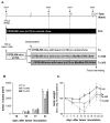
Figure 1
Figure 1
Study design for inducing melanoma tumor growth followed by monitoring cancer cachexia (A). Thirty mice were injected with 5x10⁵ melanoma cells (B16F10 cell line). As controls, ten mice were injected with RPMI medium only without tumor cells (con). The B16F10 inoculated mice were randomly assigned on day 3 to study groups either receiving standard diet during melanoma outgrowth (Tu), or received food supplemented with Myomed-946 (Tu-946), or receiving chow supplemented with Myomed-205 (Tu-205), respectively. Wire hang tests (WHTs) were performed at days 9, 16, and 23. Mice were sacrificed at day 24 for dissections. Tumor growth was monitored non-invasively by caliper measurements (B) and animal weights (C) were assessed. Values are shown as mean ± standard error of the mean, * p < 0.05 vs. Tu at the same time point, § p < 0.05 vs. con at the same time point.
Figure 2
Figure 2
Effect of Myomed-946 and -205 on skeletal muscle weights and wire hang test (WHT) expressed as maximal holding impulses in Tu mice. WHTs indicated progressively falling holding impulses until day 23 in the Tu group. Feeding with Myomed-946 and -205 attenuated this (A). Soleus (B) tibialis anterior (C) and EDL (D) muscle weights were determined at day 24 when dissecting the four study groups. Tumor outgrowth lowered muscle weights in all three skeletal muscles types. Feeding with Myomed-946 and -205 attenuated muscle wasting. Values are shown as mean ± standard error of the mean, * p < 0.05, ** p < 0.01 *** p < 0.001 vs. con; § p < 0.05, §§ p < 0.01 vs. Tu.
Figure 3
Figure 3
Effects of tumor outgrowth and feeding of Myomed-946 and -205 on heart (A), kidney (B), thymus (C), epididymal fat (D), and spleen weight (E) by day 24. Tumor outgrowth caused severe loss of epididymal fat, thymus atrophy, and splenomegaly. These effects were significantly offset by Myomed-946 or -205 feeding. Kidney and cardiac atrophy during tumor outgrowth were not significantly prevented by the compounds and remained on Tu levels below con. Clinical chemistry detected severe anemia in Tu mice as apparent by low hemoglobin (Hb) content (F). Values are shown as mean ± standard error of the mean, * p < 0.05, ** p < 0.01, *** p < 0.001 vs. con; § p < 0.05 vs. Tu.
Figure 4
Figure 4
Citrate synthase and complex I activity in groups #1-4 (A). Melanoma outgrowth leads to depleted CS and complex 1 activity. Feeding with Myomed-946 (TU-946) or Myomed-205 (TU-205) reverses this reduction (B). Values are shown as mean ± standard error of the mean, * p < 0.05 vs. con; §§§ p < 0.001 vs. Tu.
Figure 5
Figure 5
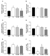
Western blot analysis were performed in the EDL muscle in all groups using specific antibodies against MuRF1 (A), MuRF2 (B), ubiquitin modified proteins (C), Nox2 (D), nitrotyrosin modified proteins (E) and LC3 (F). Values are shown as mean ± standard error of the mean. * p < 0.05 vs. con; § p < 0.05, §§ p < 0.01 vs. Tu; # p < 0.05 vs Tu-946.
Figure 6
Figure 6
Effects of 24-day Myomed-205 feeding on healthy control mice. Supplementation of food with 1g Myomed-205 per kg standard diet did not affect food intake over 24 days (A). The Myomed-205 fed mice (con-205) had further elevated body weight (~6%) at day 24 when compared to control mice (con). Note that significant differences emerged at day 10 (B). Myomed-205 feeding increased total body fat by approximately 17% (C), total lean mass by 6% (D) and maximum muscle force by 50% (E) at day 24. Values are shown as mean ± standard error of the mean, * p < 0.05 vs. con.

References

    1. Tisdale M.J. Cachexia in cancer patients. Nat. Rev. Cancer. 2002;11:862–871. doi: 10.1038/nrc927. - DOI - PubMed
    1. Argilés J.M., Busquets S., Stemmler B., López-Soriano F.J. Cancer cachexia: Understanding the molecular basis. Nat. Rev. Cancer. 2014;14:754–762. doi: 10.1038/nrc3829. - DOI - PubMed
    1. Baracos V.E., Martin L., Korc M., Guttridge D.C., Fearon K.C.H. Cancer-associated cachexia. Nat. Rev. Dis. Primers. 2018;4:17105. - PubMed
    1. Sadeghi M., Keshavarz-Fathi M., Baracos V., Arends J., Mahmoudi M., Rezaei N. Cancer cachexia: Diagnosis, assessment, and treatment. Crit. Rev. Oncol. Hematol. 2018;127:91–104. doi: 10.1016/j.critrevonc.2018.05.006. - DOI - PubMed
    1. Guglielmi V., Nowis D., Tinelli M., Malatesta M., Paoli L., Marini M., Manganotti P., Sadowski R., Wilczynski G.M., Meneghini V., et al. Bortezomib-Induced Muscle Toxicity in Multiple Myeloma. J. Neuropathol. Exp. Neurol. 2017;76:620–630. doi: 10.1093/jnen/nlx043. - DOI - PubMed

Publication types

MeSH terms