Comparative Study
. 2021 Jan;79(1):20-29.
doi: 10.1016/j.eururo.2020.09.043. Epub 2020 Oct 10.

False Positive Multiparametric Magnetic Resonance Imaging Phenotypes in the Biopsy-naïve Prostate: Are They Distinct from Significant Cancer-associated Lesions? Lessons from PROMIS

Affiliations
Comparative Study

False Positive Multiparametric Magnetic Resonance Imaging Phenotypes in the Biopsy-naïve Prostate: Are They Distinct from Significant Cancer-associated Lesions? Lessons from PROMIS

Vasilis Stavrinides et al. Eur Urol. 2021 Jan.

Abstract

Background: False positive multiparametric magnetic resonance imaging (mpMRI) phenotypes prompt unnecessary biopsies. The Prostate MRI Imaging Study (PROMIS) provides a unique opportunity to explore such phenotypes in biopsy-naïve men with raised prostate-specific antigen (PSA) and suspected cancer.

Objective: To compare mpMRI lesions in men with/without significant cancer on transperineal mapping biopsy (TPM).

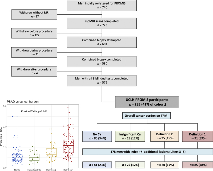
Design, setting, and participants: PROMIS participants (n=235) underwent mpMRI followed by a combined biopsy procedure at University College London Hospital, including 5-mm TPM as the reference standard. Patients were divided into four mutually exclusive groups according to TPM findings: (1) no cancer, (2) insignificant cancer, (3) definition 2 significant cancer (Gleason ≥3+4 of any length and/or maximum cancer core length ≥4mm of any grade), and (4) definition 1 significant cancer (Gleason ≥4+3 of any length and/or maximum cancer core length ≥6mm of any grade).

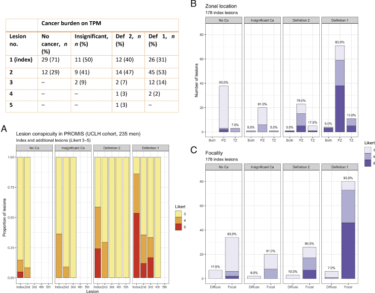
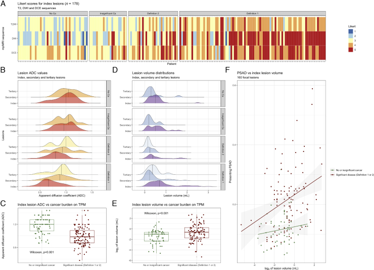
Outcome measurements and statistical analysis: Index and/or additional lesions present in 178 participants were compared between TPM groups in terms of number, conspicuity, volume, location, and radiological characteristics.

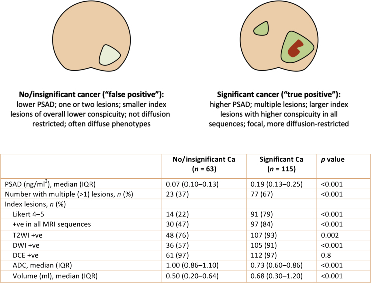
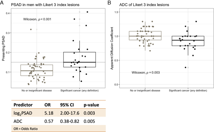
Results and limitations: Most lesions were located in the peripheral zone. More men with significant cancer had two or more lesions than those without significant disease (67% vs 37%; p< 0.001). In the former group, index lesions were larger (mean volume 0.68 vs 0.50 ml; p< 0.001, Wilcoxon test), more conspicuous (Likert 4-5: 79% vs 22%; p< 0.001), and diffusion restricted (mean apparent diffusion coefficient [ADC]: 0.73 vs 0.86; p< 0.001, Wilcoxon test). In men with Likert 3 index lesions, log2PSA density and index lesion ADC were significant predictors of definition 1/2 disease in a logistic regression model (mean cross-validated area under the receiver-operator characteristic curve: 0.77 [95% confidence interval: 0.67-0.87]).

Conclusions: Significant cancer-associated MRI lesions in biopsy-naïve men have clinical-radiological differences, with lesions seen in prostates without significant disease. MRI-calculated PSA density and ADC could predict significant cancer in those with indeterminate MRI phenotypes.

Patient summary: Magnetic resonance imaging (MRI) lesions that mimic prostate cancer but are, in fact, benign prompt unnecessary biopsies in thousands of men with raised prostate-specific antigen. In this study we found that, on closer look, such false positive lesions have different features from cancerous ones. This means that doctors could potentially develop better tools to identify cancer on MRI and spare some patients from unnecessary biopsies.

Keywords: False positive lesions; Multiparametric magnetic resonance imaging; PROMIS; Prostate cancer.


Figures

Fig. 1
Fig. 1
UCLH PROMIS cohort (n = 235). Men were classified according to mpMRI findings (Likert scores) and TPM biopsy results to four categories, as shown in the organogram: (1) no cancer, (2) insignificant cancer, (3) definition 1 cancer, and (4) definition 2 cancer. There was a significant difference in presenting PSAD between the four TPM groups (p < 0.001, Kruskal-Wallis ANOVA). In total, 178 out of 235 men (76%) had at least one lesion in their prostate and were the focus of subsequent analyses. ANOVA = analysis of variance; Ca = cancer; mpMRI = multiparametric magnetic resonance imaging; PROMIS = Prostate MRI Imaging Study; PSAD = prostate-specific antigen density; TPM = transperineal mapping biopsy; UCLH = University College London Hospital.
Fig. 2
Fig. 2
MRI lesion count, conspicuity, distribution, and focality in the UCLH PROMIS cohort. More men with any significant cancer had more than one lesion (77/115 or 67%) than those without significant disease (23/63 or 37%, p <  0.001; see the table). (A) The proportion of index lesions with high conspicuity (Likert score 4–5) increased with significant cancer burden and was significantly higher in men with significant cancer (91/115, 79%) than in those without significant disease (14/63, 22%; p <  0.001). (B) Index lesions were predominantly distributed in the PZ across all groups. (C) MRI lesions characterised by the uroradiologist as “diffuse” were more common in the TPM-negative/insignificant disease groups collectively than in men with significant disease, although this difference was not statistically significant. Ca = cancer; MRI = magnetic resonance imaging; PROMIS = Prostate MRI Imaging Study; PZ = peripheral zone; TPM= transperineal mapping biopsy; TZ = transition zone.
Fig. 3
Fig. 3
Basic radiomic characteristics of mpMRI lesions across TPM groups in the UCLH PROMIS cohort. (A) As the burden of significant disease on TPM increased, so did Likert scores for all mpMRI sequences; however, there was also a gradual shift from predominantly T2W + DWI–DCE + MRI phenotypes in men without significant cancer to T2W++DWI+++DCE++ lesions in those with significant disease. (B and C) Lesion ADC values decreased with increasing disease burden (ADC distributions shown; two outliers removed for visualisation purposes), but this reduction was particularly marked for index lesions. (D and E) Lesion volumes were greater and more skewed in men with definition 1 disease (volume distributions shown; seven outliers removed for visualisation purposes), particularly in the case of index lesions. (F) There was a positive relationship between lesion volume and PSAD, especially in men with significant cancer (regression lines shown for men with/without significant disease). ADC = apparent diffusion coefficient; Ca = cancer; DCE = dynamic contrast enhanced; DWI = diffusion-weighted imaging; mpMRI = multiparametric MRI; MRI = magnetic resonance imaging; PROMIS = Prostate MRI Imaging Study; PSAD = prostate-specific antigen density; TPM = transperineal mapping biopsy; T2W = T2 weighted.
Fig. 4
Fig. 4
Summary of the main clinical-radiological differences between men with and without significant cancer. False and true positives differ in terms of their overall PSAD level, presence or absence of additional MRI lesions, and features of index lesions (such as overall conspicuity, diffusion restriction, and volume). ADC = apparent diffusion coefficient; Ca = cancer; DCE = dynamic contrast enhanced; DWI = diffusion-weighted imaging; IQR = interquartile range; MRI = magnetic resonance imaging; PSAD = prostate-specific antigen density; T2WI = T2-weighted imaging.
Fig. 5
Fig. 5
Detection of significant cancer in men with Likert 3 index lesions. There was a significant difference in (A) PSAD and (B) index lesion ADC values between men with significant cancer and those without (PSAD threshold of 0.015 shown in grey). Both log2PSAD and index lesion ADC were significant predictors of definition 1 or 2 cancer in a binary logistic regression model (refer to the table; odds ratios shown for every PSAD doubling and for every 0.1 [ie, 100 × 10–6 mm2/s] increase in ADC). The 10-fold cross-validated mean AUC for the model was 0.77 (95% CI: 0.67–0.87). ADC = apparent diffusion coefficient; AUC = area under the receiver-operator characteristic curve; CI = confidence interval; OR = odds ratio; PSAD = prostate-specific antigen density.
Fig. 6
Fig. 6
False positive mpMRI phenotypes in TPM-negative men (n = 77; three excluded due to incomplete DICOM data). Prostate outlines, transition zones, and all Likert 3–5 lesions (41 index and 12 additional) were annotated on the Osirix platform (T2W, ADC, and DCE sequences; all axial slices). (A) Lesions in each positive sequence are superimposed on a “mean prostate shape” for the TPM-negative group, as described in the Methods. The produced maps confirm a PZ distribution and dominance of a T2W + DWI–DCE + phenotype. (B) Index lesions could broadly be divided into three morphological categories: focal (n = 34), diffuse homogeneous (n = 4), and diffuse heterogeneous (n = 3). Typical examples of lesions of each category are shown in T2W, ADC, b1400, and DCE sequences (white arrows), along with a diagrammatic representation of the three types. ADC = apparent diffusion coefficient; DCE = dynamic contrast enhanced; DWI = diffusion-weighted imaging; mpMRI = multiparametric magnetic resonance imaging; PZ = peripheral zone; TPM = transperineal mapping biopsy; T2WI = T2-weighted imaging.

Comment in

References

    1. Kasivisvanathan V., Rannikko A.S., Borghi M. MRI-targeted or standard biopsy for prostate-cancer diagnosis. N Engl J Med. 2018;378:1767–1777. - PMC - PubMed
    1. Schoots I.G. MRI in early prostate cancer detection: how to manage indeterminate or equivocal PI-RADS 3 lesions? Transl Androl Urol. 2018;7:70–82. - PMC - PubMed
    1. National Institute for Health and Care Excellence (NICE). Guidance NG 131: prostate cancer diagnosis and management. https://www.nice.org.uk/guidance/ng131. - PubMed
    1. National Prostate Cancer Audit . 2019. National prostate Cancer audit annual report.https://www.npca.org.uk/reports/npca-annual-report-2019/
    1. Drost F.-J.H., Osses D., Nieboer D. Prostate magnetic resonance imaging, with or without magnetic resonance imaging-targeted biopsy, and systematic biopsy for detecting prostate cancer: a Cochrane systematic review and meta-analysis. Eur Urol. 2020;77:78–94. - PubMed

Publication types

Substances