. 2020 Dec 4;295(49):16470-16486.
doi: 10.1074/jbc.RA120.015444. Epub 2020 Oct 13.

Co-crystal structures of HIV TAR RNA bound to lab-evolved proteins show key roles for arginine relevant to the design of cyclic peptide TAR inhibitors

Affiliations

Co-crystal structures of HIV TAR RNA bound to lab-evolved proteins show key roles for arginine relevant to the design of cyclic peptide TAR inhibitors

Sai Shashank Chavali et al. J Biol Chem. .

Abstract

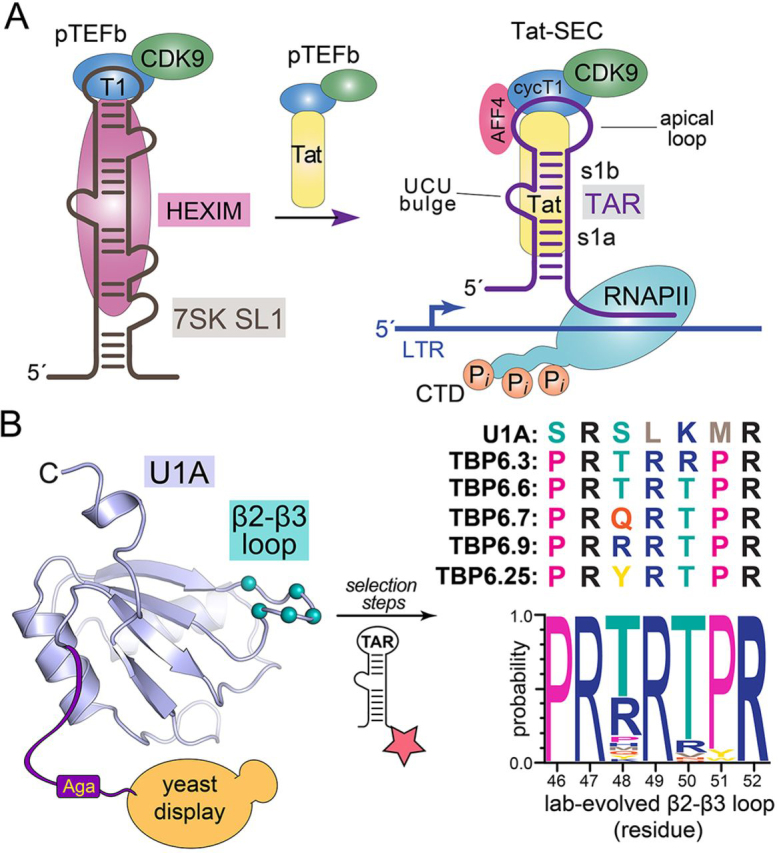
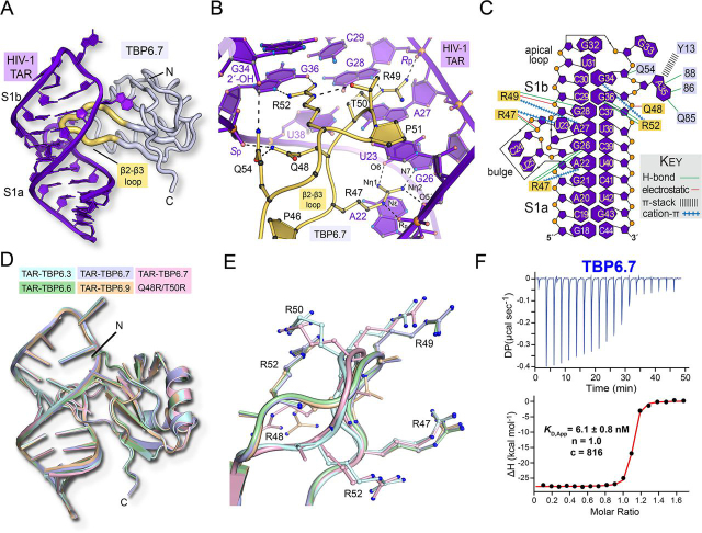
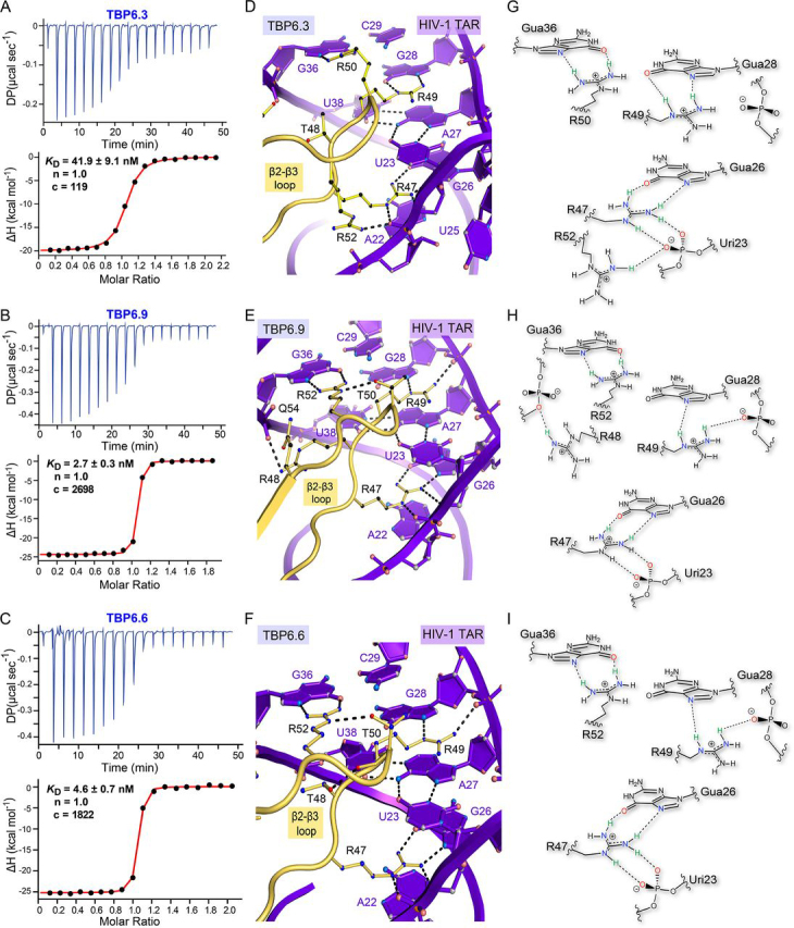
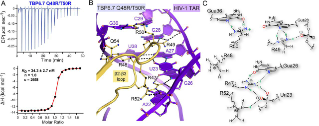
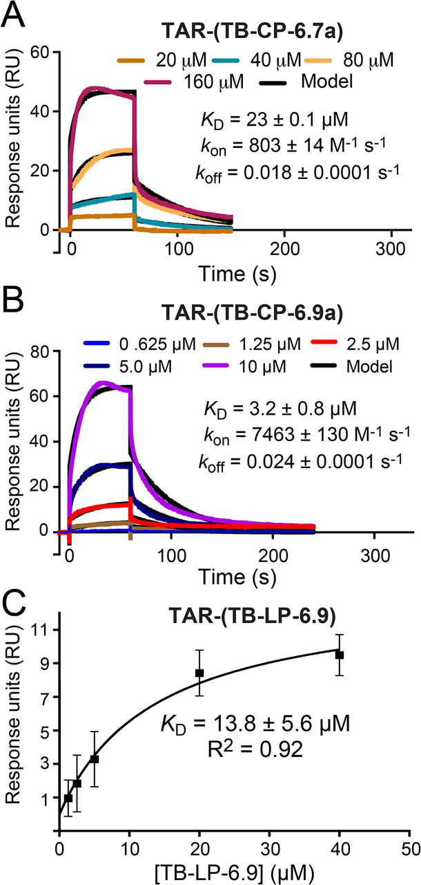
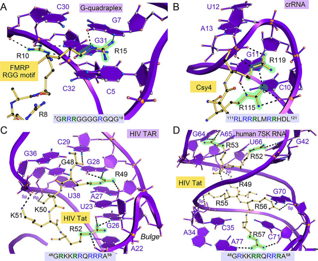
RNA-protein interfaces control key replication events during the HIV-1 life cycle. The viral trans-activator of transcription (Tat) protein uses an archetypal arginine-rich motif (ARM) to recruit the host positive transcription elongation factor b (pTEFb) complex onto the viral trans-activation response (TAR) RNA, leading to activation of HIV transcription. Efforts to block this interaction have stimulated production of biologics designed to disrupt this essential RNA-protein interface. Here, we present four co-crystal structures of lab-evolved TAR-binding proteins (TBPs) in complex with HIV-1 TAR. Our results reveal that high-affinity binding requires a distinct sequence and spacing of arginines within a specific β2-β3 hairpin loop that arose during selection. Although loops with as many as five arginines were analyzed, only three arginines could bind simultaneously with major-groove guanines. Amino acids that promote backbone interactions within the β2-β3 loop were also observed to be important for high-affinity interactions. Based on structural and affinity analyses, we designed two cyclic peptide mimics of the TAR-binding β2-β3 loop sequences present in two high-affinity TBPs (KD values of 4.2 ± 0.3 and 3.0 ± 0.3 nm). Our efforts yielded low-molecular weight compounds that bind TAR with low micromolar affinity (KD values ranging from 3.6 to 22 μm). Significantly, one cyclic compound within this series blocked binding of the Tat-ARM peptide to TAR in solution assays, whereas its linear counterpart did not. Overall, this work provides insight into protein-mediated TAR recognition and lays the ground for the development of cyclic peptide inhibitors of a vital HIV-1 RNA-protein interaction.

Keywords: HIV TAR; HIV Tat; RNA structure; RNA-binding protein; RNA-protein interaction; RNA-protein interactions; X-ray crystallography; arginine-rich domain; cyclic peptide; cyclic peptide inhibitor; drug discovery; human immunodeficiency virus (HIV); isothermal titration calorimetry; isothermal titration calorimetry (ITC); peptide chemical synthesis; surface plasmon resonance; surface plasmon resonance (SPR).


Conflict of interest statement

Conflict of interest—The authors declare that they have no conflicts of interest with the contents of this article.

Figures

Figure 1
Figure 1
Schematic diagram of Tat interactions with human 7SK and HIV TAR leading to the SEC, and overview of the selection process that produced lab-evolved TBPs.A, left, cartoon depicting the inactive pTEFb complex sequestered by the host 7SK small-nuclear ribonucleoprotein (7SK RNP) complex that includes protein HEXIM. The ARM of HIV-1 Tat protein mimics that of HEXIM, displacing the pTEFb complex, which contains CycT1 and CDK9. Right arrow, Tat transfers the pTEFb complex by direct interaction with HIV-1 TAR at the 5′-LTR. This complex subsequently forms the SEC. Transcription elongation of viral mRNA is then activated by CDK9-mediated phosphorylation of the RNA polymerase II C-terminal domain (6, 7). B, left, crystal structure of U1A from the protein-RNA complex (PDB entry 1URN) showing amino acids that were diversified in the β2-β3 loop based on their contact with hpII RNA (8). Selection was performed by yeast display and cell sorting based on binding to fluorescently labeled TAR (star). The U1A fold exhibits a βαββαβα topology typical of the single-stranded RRM (9). Right, sequences of the β2-β3 loop from U1A and lab-evolved TBPs of this investigation. A Weblogo analysis highlights the consensus of the lab-evolved β2-β3 loop. Adapted from Ref. .
Figure 2
Figure 2
Structural overview of lab-evolved protein TBP6.7, superpositions of HIV TAR-TBP co-crystal structures of this investigation, and TAR binding to TBP6.7.A, ribbon diagram depicting the overall fold of the TAR-TBP6.7 complex reveals entry of the β2-β3 loop into the major groove (PDB entry 6CMN) (28). B, close-up view of the lab-evolved β2-β3 loop of TBP6.7 showing TAR readout that includes Arg-47, which reads the Hoogsteen edge of Gua26 and the Uri23 backbone; Arg-49, which recognizes N7 of Gua28 and the phosphate backbone; and Arg-52, which reads the Hoogsteen edge of Gua36. Here and elsewhere, putative interactions are shown by dashed lines. C, overview of chemical interactions between the TAR-TBP6.7 complex that are representative of the various RNA-protein interactions of other TBPs in the current investigation. D, pairwise all-atom superposition of the co-crystal structures of this investigation upon the TAR-TBP6.7 complex. The average r.m.s.d. was 0.43 Å. The overall three-dimensional fold of each TBP is similar to TBP6.7. TAR RNA also reveals structurally similar details (Fig. S2). E, close-up view of the superimposed β2-β3 loops from D. Arginine placement affects the loop conformation and the mode of TAR recognition. The average loop r.m.s.d. was 0.78 Å. F, representative ITC thermogram of TBP6.7 titrated into TAR. The apparent equilibrium dissociation constant (KD) is shown, along with the stoichiometry (n), and the c value to indicate the quality of the binding model fit (34). Here and elsewhere, the representative single-run ITC parameters shown on thermograms differ from Table 2, which reports average values from duplicate titrations.
Figure 3
Figure 3
Affinity analysis of TBP variants for HIV-1 TAR and close-up views of TAR-TBP co-crystal structures revealing arginine-mediated RNA recognition.A, representative ITC thermogram of TBP6.3 titrated into TAR. B, representative ITC thermogram of TBP6.9 titrated into TAR. C, representative ITC thermogram of TBP6.6 titrated into TAR. D, close-up view of the lab-evolved β2-β3 loop of TBP6.3 showing TAR readout by four arginines. Arg-47 and Arg-49 retain interactions similar to TBP6.7 (Fig. 2, B and C), but Arg-52 is displaced by Arg-50 to recognize Gua36. As a result, Arg-52 now binds the backbone pro-Rp oxygen of Uri23. E, close-up view of the lab-evolved β2-β3 loop of TBP6.9 showing TAR readout by four arginines, including three consecutive arginines. Arg-48 interacts with the backbone at the pro-Sp oxygen of Gua36, similar to Gln-48 of TBP6.7, whereas Arg-47, Arg-49, and Arg-52 retain Hoogsteen-edge readout similar to TBP6.7 (Fig. 2, B and C). F, close-up view of the lab-evolved β2-β3 loop of TBP6.6 showing TAR readout by three arginines. The mode of interaction is comparable with TBP6.7 (Fig. 2B), except that Thr-48 and Thr-50 engage in stabilizing side chain-to-backbone interactions. G, schematic diagram depicting arginine interactions between TBP6.3 and TAR in D. H, schematic diagram depicting arginine interactions between TBP6.9 and TAR in E. I, schematic diagram depicting arginine interactions between TBP6.6 and TAR in F.
Figure 4
Figure 4
Affinity analysis of five-arginine mutant TBP6.7 Q48R/T50R for TAR RNA and close-up view of the corresponding co-crystal structure.A, representative thermogram of TBP6.7 Q48R/T50R titrated into TAR. B, close-up view of variant TBP6.7 Q48R/T50R showing readout of TAR using four arginines. Arg-47 retains interactions similar to TBP6.7 (Fig. 2, B and C). Arg-48 interacts with the pro-Sp nonbridging oxygen of Gua36, as seen for TBP6.9 (Fig. 3, E and H). Nε and of Nη2 of Arg-49 contact N7 and the pro-Rp oxygen of Gua28. Arg-50 recognizes the Hoogsteen edge of Gua36, displacing Arg-52, which salt-bridges to the pro-Rp oxygen of Uri23. The former and latter interactions parallel Arg-50 of TBP6.3 and Arg-52 of TBP6.9 (Fig. 3 (panels d and g and panels e and h)). C, schematic diagram depicting arginine interactions from B and their interactions in the co-crystal structure. Like all other TBPs, a trio of arginines recognizes the Hoogsteen edges of conserved guanines in the major groove. There are no apparent intrapeptide stabilization interactions.
Figure 5
Figure 5
Kinetic and equilibrium binding analysis of peptides TB-CP-6.7a, TB-CP-6.9a, and TB-LP-6.9 binding to HIV-1 TAR.A, representative sensorgrams from SPR showing cyclic peptide TB-CP-6.7a association with and dissociation from immobilized TAR RNA. Here and elsewhere, peptide concentrations are shown in the key; colored lines represent background-subtracted data; black lines indicate the global fit to a 1:1 binding model. The binding parameters obtained from the data set are shown; the χ2 (RU2) quality control metric for the fit was 0.76. For this and other experiments, average kon and koff rate constants and the apparent KD value from replicate runs are reported in Table 3. B, representative sensorgrams from SPR showing cyclic peptide TB-CP-6.9a association with and dissociation from immobilized TAR RNA. This experiment was conducted in the presence of a 100-fold molar excess tRNA. The χ2 (RU2) for the fit was 1.95. C, equilibrium binding analysis of linear peptide TB-LP-6.9 interacting with TAR; the average KD is shown.
Figure 6
Figure 6
Cyclic peptide competition with the Tat ARM for HIV TAR binding.A, schematic diagram of the TAR-Tat competition experiment wherein cyclic peptide TB-CP-6.9a is titrated initially into TAR RNA. The Tat ARM is titrated subsequently into the preformed complex. In a successful assay, the presence of TB-CP-6.9a competes with the Tat ARM to block the Tat-TAR interaction. The TAR structure used for this schematic was derived from the TAR-TBP6.9 complex, and the Tat ARM structure was derived from the lowest-energy conformation of the Tat-TAR complex (PDB 6MCE) (11). B, representative control titration of the Tat ARM peptide into HIV-1 TAR RNA produced an average KD of 135 ± 31 nm and with 2:1 stoichiometry, suggesting two high-affinity binding sites. For this and other experiments, the values indicated in each ITC panel correspond to individual thermograms, whereas average values are provided in Table 2. C, representative titration of cyclic peptide TB-CP-6.9a binding to TAR, which yielded an average KD of 5.3 ± 0.2 μm with 1:1 binding stoichiometry. D, representative competition titration in which the Tat ARM peptide was titrated into the product formed in C. The binding reaction shows no appreciable heats of binding. Each experiment in B–D was performed twice.
Figure 7
Figure 7
Close-up views depicting how various proteins containing ARMs use only a subset of arginines to recognize their cognate RNAs.A, close-up view of the FMRP-RGG-RNA interface (PDB entry 5DEA) (51) (boxed area in Fig. S6A). The β-hairpin RGG motif contains four arginines (sequence shown), but only two make base-specific contacts (highlighted green). Arg-10 and Arg-15 recognize the Hoogsteen edges of Gua31 and Gua7, but Arg-8 and Arg-9 do not make base-specific interactions. B, close-up view of the Csy4 endoribonuclease in complex with crRNA (PDB entry 4AL5) (52) (boxed area in Fig. S6B). The α-helical motif harbors six arginines, but only two participate in binding. Arg-115 reads the Hoogsteen edge of Gua11, and Arg-119 makes phosphate backbone interactions. Other arginines are either involved in salt-bridge interactions with the RNA backbone or do not make RNA contacts. C, close-up view of the HIV TAR-Tat interface (PDB entry 6MCE) (19) (boxed area in Fig. S6C). The ARM contains nine arginines, but only two make base-specific interactions. Arg-49 hydrogen-bonds to the Hoogsteen edge of Gua28. Similarly, Arg-52 hydrogen-bonds with the Hoogsteen edge of Gua26. Other arginines do not make base-specific interactions. D, close-up view of the HIV Tat-7SK RNA interface (PDB entry 6MCF) (11) (boxed area in Fig. S6D). The Tat ARM with nine arginines utilizes only three to make base-specific interactions. Specifically, Arg-52, Arg-53, and Arg-57 recognize the Hoogsteen edges of Gua42, Gua64, and Ade77. Arg-49, Arg-55, and Arg-56 do not make base-specific interactions.

References

    1. Gerstberger S., Hafner M., Tuschl T. A census of human RNA-binding proteins. Nat. Rev. Genet. 2014;15:829–845. doi: 10.1038/nrg3813. 25365966. - DOI - PMC - PubMed
    1. He C., Sidoli S., Warneford-Thomson R., Tatomer D.C., Wilusz J.E., Garcia B.A., Bonasio R. High-resolution mapping of RNA-binding regions in the nuclear proteome of embryonic stem cells. Mol. Cell. 2016;64:416–430. doi: 10.1016/j.molcel.2016.09.034. 27768875. - DOI - PMC - PubMed
    1. Zagrovic B., Bartonek L., Polyansky A.A. RNA-protein interactions in an unstructured context. FEBS Lett. 2018;592:2901–2916. doi: 10.1002/1873-3468.13116. 29851074. - DOI - PMC - PubMed
    1. Bell N.M., L'Hernault A., Murat P., Richards J.E., Lever A.M., Balasubramanian S. Targeting RNA-protein interactions within the human immunodeficiency virus type 1 lifecycle. Biochemistry. 2013;52:9269–9274. doi: 10.1021/bi401270d. 24358934. - DOI - PMC - PubMed
    1. Engelman A., Cherepanov P. The structural biology of HIV-1: mechanistic and therapeutic insights. Nat. Rev. Microbiol. 2012;10:279–290. doi: 10.1038/nrmicro2747. 22421880. - DOI - PMC - PubMed

Uncited reference

    1. Hodel A., Kim S.H., Brunger A.T. Model bias in macromolecular crystal-structures. Acta Crystallogr. A. 1992;48:851–858. doi: 10.1107/S0108767392006044. - DOI
    1. Brunger A.T., Adams P.D., Clore G.M., DeLano W.L., Gros P., Grosse-Kunstleve R.W., Jiang J.S., Kuszewski J., Nilges M., Pannu N.S., Read R.J., Rice L.M., Simonson T., Warren G.L. Crystallography & NMR system: a new software suite for macromolecular structure determination. Acta Crystallogr. D Biol. Crystallogr. 1998;54:905–921. doi: 10.1107/s0907444998003254. 9757107. - DOI - PubMed

Publication types

MeSH terms

LinkOut - more resources