. 2020 Oct 27;117(43):26795-26803.
doi: 10.1073/pnas.2008830117. Epub 2020 Oct 13.

p31comet promotes homologous recombination by inactivating REV7 through the TRIP13 ATPase

Affiliations

p31comet promotes homologous recombination by inactivating REV7 through the TRIP13 ATPase

Prabha Sarangi et al. Proc Natl Acad Sci U S A. .

Abstract

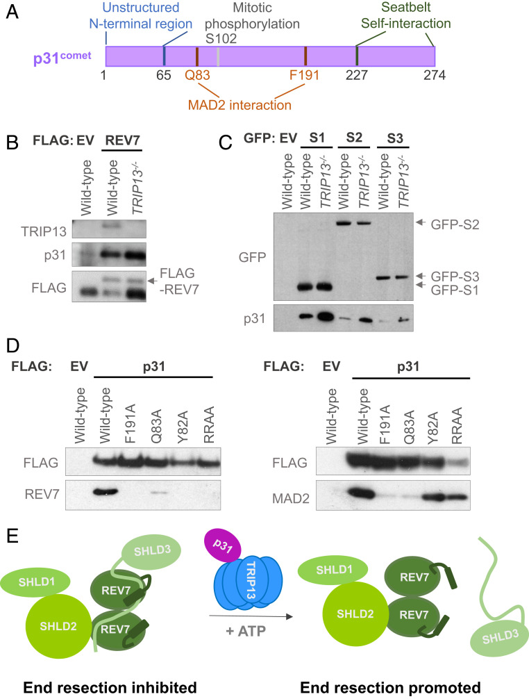
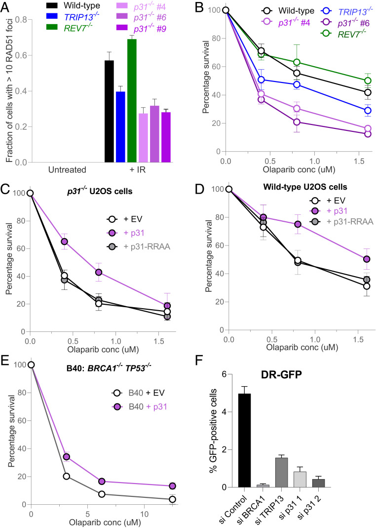
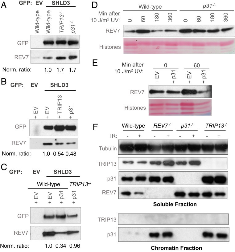
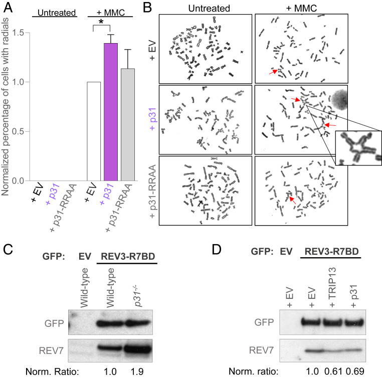
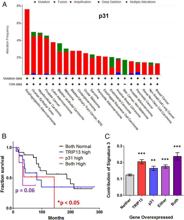
The repair of DNA double strand breaks (DSBs) that arise from external mutagenic agents and routine cellular processes is essential for life. DSBs are repaired by two major pathways, homologous recombination (HR) and classical nonhomologous end joining (C-NHEJ). DSB repair pathway choice is largely dictated at the step of 5'-3' DNA end resection, which is promoted during S phase, in part by BRCA1. Opposing end resection is the 53BP1 protein, which recruits the ssDNA-binding REV7-Shieldin complex to favor C-NHEJ repair. We recently identified TRIP13 as a proresection factor that remodels REV7, causing its dissociation from the Shieldin subunit SHLD3. Here, we identify p31comet, a negative regulator of MAD2 and the spindle assembly checkpoint, as an important mediator of the TRIP13-REV7 interaction. p31comet binds to the REV7-Shieldin complex in cells, promotes REV7 inactivation, and causes PARP inhibitor resistance. p31comet also participates in the extraction of REV7 from the chromatin. Furthermore, p31comet can counteract REV7 function in translesion synthesis (TLS) by releasing it from REV3 in the Pol ζ complex. Finally, p31comet, like TRIP13, is overexpressed in many cancers and this correlates with poor prognosis. Thus, we reveal a key player in the regulation of HR and TLS with significant clinical implications.

Keywords: Fanconi anemia; PARP inhibitor; REV7; homologous recombination; translesion synthesis.


Conflict of interest statement

Competing interest statement: A.D.D. is a consultant/advisory board member for Lilly Oncology, Merck-EMD Serono, Intellia Therapeutics, Sierra Oncology, Cyteir Therapeutics, Third Rock Ventures, AstraZeneca, Ideaya Inc., Cedilla Therapeutics Inc., a stockholder in Ideaya Inc., Cedilla Therapeutics Inc., and Cyteir, and reports receiving commercial research grants from Lilly Oncology and Merck-EMD Serono.

Figures

Fig. 1.
Fig. 1.
p31 physically interacts with REV7-Shieldin. (A) Schematic of p31 protein showing its various functional regions and modification sites. (B) Western blot showing FLAG immunoprecipitation of FLAG-empty vector (EV) or FLAG-REV7 in wild-type and TRIP13−/− U2OS cells and the coimmunoprecipitation of endogenous p31. (C) Western blot showing GFP immunoprecipitation of GFP-tagged: empty vector (EV), SHLD1 (S1), SHLD2 (S2), and SHLD3 (S3) in wild-type and TRIP13−/− U2OS cells and the coimmunoprecipitation of endogenous p31. (D) Western blot showing the FLAG immunoprecipitation of EV, wild-type p31, and various mutant forms of p31, and the coimmunoprecipitation of endogenous REV7 (Left) or MAD2 (Right) in p31−/− HEK293T cells. RRAA denotes the RR188,189AA mutant. (E) Schematic of our proposed model of p31 function as mediating the interaction between REV7 and TRIP13, and hence the dissolution of REV7-Shieldin, through the remodeling of the REV7 seatbelt that is bound to SHLD3. Here, we depict the seatbelts of both REV7 monomers as being unlatched by the action of TRIP13-p31, but it is possible that only the seatbelt closed over SHLD3 is opened.
Fig. 2.
Fig. 2.
p31 promotes homologous recombination. (A) Graph showing RAD51 focus formation in wild-type, TRIP13−/−, REV7−/−, and three p31−/− clones in U2OS cells at baseline and 6 h following treatment with 5 Gy of ionizing radiation. Means and SDs of three independent experiments are depicted. (B) Clonogenic survival curve of wild type, TRIP13−/−, REV7−/−, and two p31−/− clones in U2OS cells at various doses of the PARP inhibitor, olaparib. Means and SDs of three independent experiments are depicted. (C) Clonogenic survival curve of p31−/− U2OS cells expressing empty vector (EV), wild-type p31 (WT), or RR188,189AA mutant p31 (RRAA) at various doses of olaparib. Means and SDs of three independent experiments are depicted. (D) Clonogenic survival curve of U2OS cells expressing EV or overexpressing WT or RR188,189AA (RRAA) mutant p31 at various doses of the PARP inhibitor, olaparib. Means and SDs of three independent experiments are depicted. (E) Clonogenic survival curve of BRCA1−/− TP53−/− RPE-1 cells, expressing EV or overexpressing p31. Means and SDs of two independent experiments are depicted. (F) DR-GFP assay results in U2OS cells following transfection with nontargeting or siRNAs targeting BRCA1, TRIP13, and p31. Means and SDs of two independent experiments are depicted.
Fig. 3.
Fig. 3.
p31, like TRIP13, inhibits REV7 activity. (A) Western blot showing the GFP immunoprecipitation of GFP-empty vector (EV) or GFP-SHLD3 and the coimmunoprecipitation of endogenous REV7 in wild-type, TRIP13−/−, and p31−/− U2OS cells. (B) Western blot showing the GFP immunoprecipitation of GFP-empty vector (EV) or GFP-SHLD3 and the coimmunoprecipitation of endogenous REV7 in U2OS cells ectopically overexpressing EV or cDNAs encoding TRIP13 or p31. (C) Western blot showing the GFP immunoprecipitation of GFP- EV or GFP-SHLD3 and the coimmunoprecipitation of endogenous REV7 in wild-type or TRIP13−/− U2OS cells expressing EV or overexpressing p31. (D) Western blot of chromatin-bound REV7 from wild-type or p31−/− U2OS cells at various time points following irradiation with 10 J/m2 of UV light. (E) Western blot of chromatin-bound REV7 from U2OS cells ectopically overexpressing EV or p31 cDNA following irradiation with 10 J/m2 of UV light. (F) Western blot of soluble (Top) and chromatin-bound (Bottom) proteins from wild-type, REV7−/−, p31−/−, and TRIP13−/− U2OS cells, with and without exposure to 5 Gy of ionizing radiation. Norm. ratio is the ratio of coimmunoprecipitated REV7 to the tagged protein which was pulled down, normalized to the control condition.
Fig. 4.
Fig. 4.
p31 inhibits REV7 activity in ICL repair. (A) Graph showing the proportion of HEK293T cells showing radial chromosomes following treatment with 20 ng/mL of MMC upon expression of empty vector (EV) or overexpression of p31 wild type or RRAA mutant. Asterisk denotes a statistically significant difference. (B) Representative images of metaphase chromosome spreads quantified in A. Arrows indicate radial chromosomes. (C) Western blot showing GFP immunoprecipitation of GFP- EV or GFP-REV3-R7BD (REV7-binding domain) and the coimmunoprecipitation of endogenous REV7 in wild-type and p31−/− U2OS cells. (D) Western blot showing GFP immunoprecipitation of GFP-EV or GFP-REV3-R7BD and the coimmunoprecipitation of endogenous REV7 in U2OS cells expressing EV or overexpressing TRIP13 or p31. Norm. ratio is the ratio of coimmunoprecipitated REV7 to the tagged protein which was pulled down, normalized to the control condition.
Fig. 5.
Fig. 5.
Overexpression of p31, similar to TRIP13, is commonly observed in cancers, correlates with poor prognosis, and contributes to HR mutation signature. (A) Bar chart showing the prevalence of amplifications (red), deletions (blue), and mutations (green) of the p31 gene across an array of cancer types in TCGA. (B) Kaplan–Meier survival curve of BRCA1-deficient breast cancer patients with overexpression of TRIP13, p31, or both as compared to cells with normal expression of both genes. Normal expression vs. p31 high: P < 0.05, normal expression vs. both high: P = 0.06, Mantel–Cox log-rank test. (C) Graph showing the contribution of the HR-deficiency signature 3 in breast cancers overexpressing TRIP13 and/or p31 compared to breast cancers with normal expression of both genes. **P < 0.01, ***P < 0.001, Mann–Whitney test, two tailed.

Comment in

References

    1. Chatterjee N., Walker G. C., Mechanisms of DNA damage, repair, and mutagenesis. Environ. Mol. Mutagen. 58, 235–263 (2017). - PMC - PubMed
    1. Mirman Z., de Lange T., 53BP1: A DSB escort. Genes Dev. 34, 7–23 (2020). - PMC - PubMed
    1. Ceccaldi R., Rondinelli B., D’Andrea A. D., Repair pathway choices and consequences at the double-strand break. Trends Cell Biol. 26, 52–64 (2016). - PMC - PubMed
    1. Symington L. S., Mechanism and regulation of DNA end resection in eukaryotes. Crit. Rev. Biochem. Mol. Biol. 51, 195–212 (2016). - PMC - PubMed
    1. Densham R. M., Morris J. R., Moving mountains-the BRCA1 promotion of DNA resection. Front. Mol. Biosci. 6, 79 (2019). - PMC - PubMed

Publication types

MeSH terms

LinkOut - more resources