

From People to Panthera: Natural SARS-CoV-2 Infection in Tigers and Lions at the Bronx Zoo
- PMID: 33051368
- PMCID: PMC7554670
- DOI: 10.1128/mBio.02220-20
From People to Panthera: Natural SARS-CoV-2 Infection in Tigers and Lions at the Bronx Zoo
Abstract
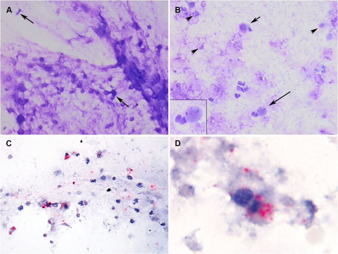
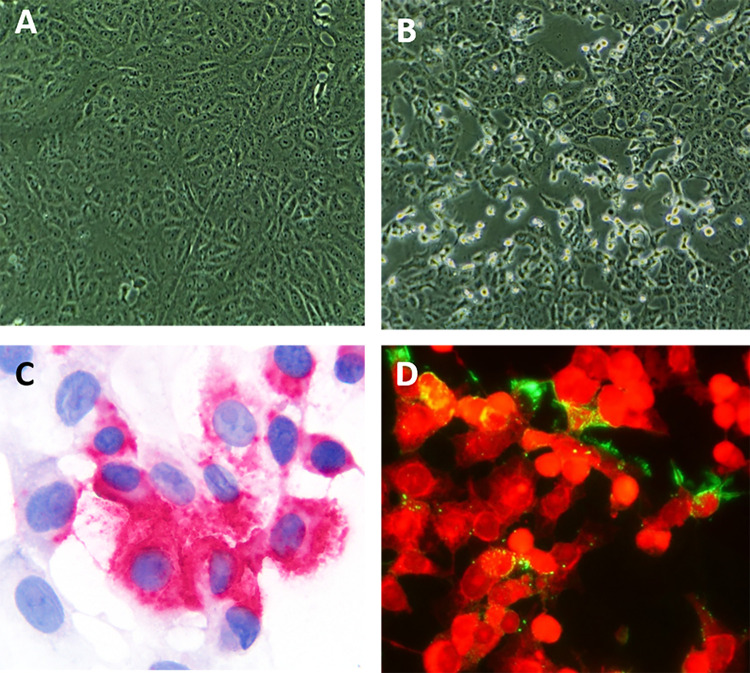
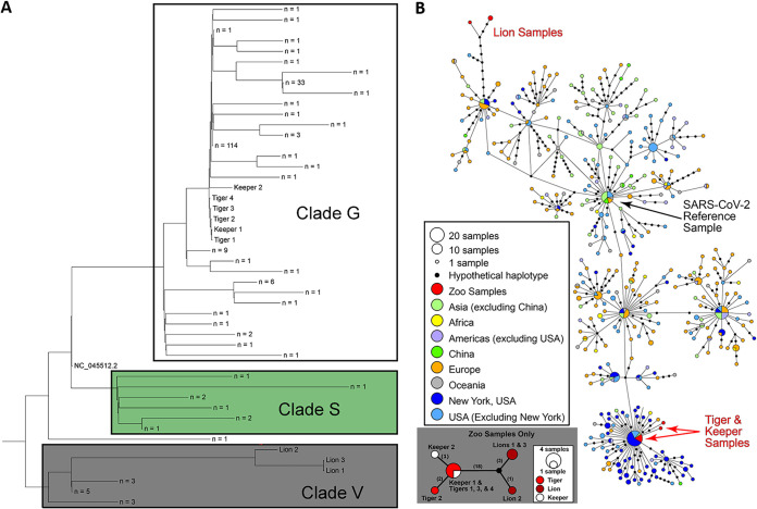
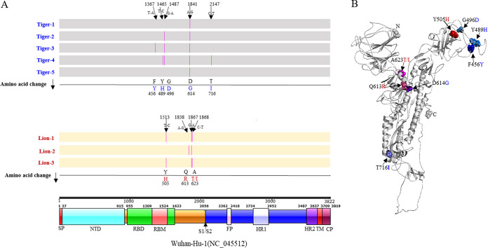
Despite numerous barriers to transmission, zoonoses are the major cause of emerging infectious diseases in humans. Among these, severe acute respiratory syndrome (SARS), Middle East respiratory syndrome (MERS), and ebolaviruses have killed thousands; the human immunodeficiency virus (HIV) has killed millions. Zoonoses and human-to-animal cross-species transmission are driven by human actions and have important management, conservation, and public health implications. The current SARS-CoV-2 pandemic, which presumably originated from an animal reservoir, has killed more than half a million people around the world and cases continue to rise. In March 2020, New York City was a global epicenter for SARS-CoV-2 infections. During this time, four tigers and three lions at the Bronx Zoo, NY, developed mild, abnormal respiratory signs. We detected SARS-CoV-2 RNA in respiratory secretions and/or feces from all seven animals, live virus in three, and colocalized viral RNA with cellular damage in one. We produced nine whole SARS-CoV-2 genomes from the animals and keepers and identified different SARS-CoV-2 genotypes in the tigers and lions. Epidemiologic and genomic data indicated human-to-tiger transmission. These were the first confirmed cases of natural SARS-CoV-2 animal infections in the United States and the first in nondomestic species in the world. We highlight disease transmission at a nontraditional interface and provide information that contributes to understanding SARS-CoV-2 transmission across species.IMPORTANCE The human-animal-environment interface of severe acute respiratory syndrome coronavirus 2 (SARS-CoV-2) is an important aspect of the coronavirus disease 2019 (COVID-19) pandemic that requires robust One Health-based investigations. Despite this, few reports describe natural infections in animals or directly link them to human infections using genomic data. In the present study, we describe the first cases of natural SARS-CoV-2 infection in tigers and lions in the United States and provide epidemiological and genetic evidence for human-to-animal transmission of the virus. Our data show that tigers and lions were infected with different genotypes of SARS-CoV-2, indicating two independent transmission events to the animals. Importantly, infected animals shed infectious virus in respiratory secretions and feces. A better understanding of the susceptibility of animal species to SARS-CoV-2 may help to elucidate transmission mechanisms and identify potential reservoirs and sources of infection that are important in both animal and human health.
Keywords: One Health; Panthera leo; Panthera tigris; SARS-CoV-2; in situ hybridization; lion; rRT-PCR; tiger; virus isolation; whole-genome sequencing; zoo; zoonotic infection.
Copyright © 2020 McAloose et al.
Figures
References
-
- Gorbalenya AE, Baker SC, Baric RS, de Groot RJ, Drosten C, Gulyaeva AA, Haagmans BL, Lauber C, Leontovich AM, Neuman BW, Penzar D, Perlman S, Poon LLM, Samborskiy DV, Sidorov IA, Sola I, Ziebuhr J. 2020. The species Severe Acute Respiratory Syndrome-related coronavirus: classifying 2019-nCoV and naming it SARS-CoV-2. Nat Microbiol 5:536–544. doi: 10.1038/s41564-020-0695-z. - DOI - PMC - PubMed
-
- Zhu N, Zhang D, Wang W, Li X, Yang B, Song J, Zhao X, Huang B, Shi W, Lu R, Niu P, Zhan F, Ma X, Wang D, Xu W, Wu G, Gao GF, Tan W, China Novel Coronavirus Investigating and Research Team. 2020. A novel coronavirus from patients with pneumonia in China, 2019. N Engl J Med 382:727–733. doi: 10.1056/NEJMoa2001017. - DOI - PMC - PubMed
-
- Zhou P, Yang XL, Wang XG, Hu B, Zhang L, Zhang W, Si HR, Zhu Y, Li B, Huang CL, Chen HD, Chen J, Luo Y, Guo H, Jiang RD, Liu MQ, Chen Y, Shen XR, Wang X, Zheng XS, Zhao K, Chen QJ, Deng F, Liu LL, Yan B, Zhan FX, Wang YY, Xiao GF, Shi ZL. 2020. A pneumonia outbreak associated with a new coronavirus of probable bat origin. Nature 579:270–273. doi: 10.1038/s41586-020-2012-7. - DOI - PMC - PubMed
Publication types
MeSH terms
Grants and funding
LinkOut - more resources
Full Text Sources
Molecular Biology Databases
Miscellaneous
